 Your shopping cart is currently empty


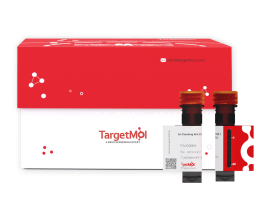
DiO Staining Solution
| Pack Size | Price | USA Warehouse | Global Warehouse | Quantity |
|---|---|---|---|---|
| 1 mL | $97 | - | In Stock |
 Product Information
| DiO Staining Solution | Specifications |
|---|---|
| Ingredient | DiO |
| CAS | 34215-57-1 |
| Conc. | 5 mM |
| Solvent | DMSO |
 Features
1.High specificity for cell membrane labeling with precise membrane localization.
2.Dye molecules align on the outer layer of the cell membrane without disrupting its structure, making it suitable for studying the membrane in its native state.
3.Low cytotoxicity: suitable for live-cell labeling studies.
4.Stable fluorescence signal: with excellent photobleaching resistance, ideal for long-term labeling.
5.Broad application: compatible with various sample types.
6.Simple operation and rapid detection.
 Application
-
Live-cell tracking (cell migration, fusion, division, and adhesion);
-
Membrane fluidity analysis;
-
Cell differentiation pathway tracing;
-
Cell-cell interaction studies;
-
Drug delivery system detection.
 Preparation of Working Solution
Dilute the DiO stock solution with an appropriate diluent (serum-free medium, PBS, or HBSS) to prepare the staining working solution (1-30 µM). The specific working concentration of DiO should be optimized according to experimental conditions; generally, a concentration of 5-10 µM is commonly used for fluorescent labeling of cell membranes.
 Instructions
Staining of Suspended Live Cells
(1) Collect suspended cells in the logarithmic growth phase and centrifuge at 1000 rpm for 5 min at room temperature. Discard the supernatant. Resuspend the cells in PBS, centrifuge again at 1000 rpm for 5 min at room temperature, and discard the supernatant.
(2) Resuspend the cells in pre-warmed (37°C) culture medium and adjust the cell density to 1×10⁶–5×10⁶ cells/mL.
(3) Add DiO working solution at one-tenth of the cell suspension volume, mix gently, and incubate at 37°C protected from light for 5–20 min.
(4) Centrifuge at 1000 rpm for 5 min and discard the supernatant. Wash the cells 2–3 times with pre-warmed (37°C) culture medium, then resuspend the cells.
(5) Analyze the cells by flow cytometry or place a drop of the cell suspension on a glass slide, cover with a coverslip, and observe under a fluorescence microscope. Excitation ≈ 484 nm; Emission ≈ 501 nm.
Staining of Adherent Live Cells
(1) Remove the culture medium and wash the cells once with pre-warmed (37°C) culture medium.
(2) Add an appropriate volume of DiO working solution to cover all cells, and incubate at 37°C protected from light for 5–20 min.
(3) Remove the DiO working solution and wash the cells 2–3 times with pre-warmed (37°C) culture medium, 5 min each time.
(4) Add pre-warmed (37°C) culture medium to cover the cells and observe under a fluorescence microscope. Excitation ≈ 484 nm; Emission ≈ 501 nm.
 Storage
Store at -20 ℃, protected from light for 12 months.
 Precautions
1.For live-cell staining with DiO, incubation at 37°C is recommended; for fixed cells or tissue samples, staining can be performed at room temperature. If fixation is required, 4% paraformaldehyde is generally recommended as the fixative.
2.During staining of suspended cells, gently shake the centrifuge tube every 10 minutes to prevent cell sedimentation and aggregation, which may affect staining uniformity.
3.Since staining efficiency can vary depending on sample type and experimental conditions, it is recommended to optimize the working solution concentration and staining time through a preliminary experiment.
4.DiO is unstable in aqueous solution and tends to aggregate over time, resulting in reduced fluorescence efficiency. Therefore, it is recommended to prepare the DiO working solution freshly before use.
5.Cells in the logarithmic growth phase have higher viability and better membrane fluidity. It is advisable to use cells in this phase for staining and avoid using aged or over-confluent cells, as impaired membrane function may affect dye binding.
6.The product is for R&D use only, not for diagnostic procedures, food, drug, household or other uses.
7.Please wear a lab coat and disposable gloves.
 Instruction Manual
| Size | Quantity | Unit Price | Amount | Operation |
|---|

Copyright © 2015-2026 TargetMol Chemicals Inc. All Rights Reserved.


