 Your shopping cart is currently empty


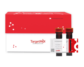
CFSE (Cell Proliferation Tracer Fluorescent Probe)
The principle of CFSE in proliferation detection and tracing is as follows: once CFSE enters viable cells, intracellular esterases specifically hydrolyze it, removing the acetate groups and generating carboxyfluorescein succinimidyl ester (CFSE). This product covalently binds to intracellular proteins via amino groups, thereby allowing CFSE to be stably retained within the cells and emit bright green fluorescence (excitation wavelength: 492 nm; emission wavelength: 517 nm).
When CFSE-labeled cells divide, the fluorescence is equally distributed between the two daughter cells, causing the fluorescence intensity of each daughter cell to be half that of the parent cell. By monitoring the changes in green fluorescence intensity, it is possible to clearly distinguish undivided cells from cells that have undergone different numbers of divisions. As cell division progresses, the gradient changes in fluorescence intensity can be used to trace the process of cell proliferation, the number of divisions, as well as cell migration and distribution both in vitro and in vivo.
| Pack Size | Price | USA Warehouse | Global Warehouse | Quantity |
|---|---|---|---|---|
| 1 mL | $129 | - | In Stock |
 Product Information
| CFSE (Cell Proliferation Tracer Fluorescent Probe) | Specifications |
|---|---|
| Ingredient | CFSE |
| CAS | 150347-59-4 |
| Conc. | 10 mM |
| Solvent | DMSO |
 Features
1.Excellent labeling performance: the intracellular fluorescence signal after staining is bright and uniform.
2.Stable fluorescence signal: the fluorescence can be maintained for several days after staining.
3.Low cytotoxicity: it does not interfere with normal physiological activities such as cell proliferation.
4.Reliable results: the fluorescent dye is transferred only to daughter cells through cell division, and not to adjacent cells by proximity.
5.Simple operation and rapid detection.
 Application
Cell proliferation assay; Cell tracking study; Exosome uptake tracing; Live-cell differentiation trajectory monitoring.
 Preparation of Working Solution
Dilute the CFSE stock solution with an appropriate diluent (serum-free medium or PBS buffer) to prepare a CFSE working solution at a concentration of 1-10 µM.
Note: The exact working concentration should be adjusted according to the experimental requirements. The working solution should be freshly prepared before use.
 Instructions
For adherent cells
(1)Seed adherent cells on sterile coverslips for culture.
(2)Once cell culture is complete, remove the old medium and wash the cells once with pre-warmed PBS (37 ℃).
(3)Add pre-warmed CFSE staining working solution (37 ℃), and incubate the cells in a 37 ℃ incubator protected from light for 15 min.
(4)Remove the staining solution, add an appropriate volume of pre-warmed culture medium (37 ℃), and incubate in a 37℃ incubator protected from light for 30 min.
(5)Remove the old medium and wash the cells with pre-warmed PBS (37 ℃) 1-2 times.
(6)Add pre-warmed culture medium (37 ℃) to cover the cells. Observe the staining results under a fluorescence microscope. Excitation wavelength can be set at 492 nm and emission wavelength at 517 nm to detect fluorescence.
Note: If flow cytometry is required for adherent cells, digest the adherent cells with trypsin, collect and resuspend them, and then follow the staining procedure for suspension cells below.
For suspension cells
(1)Centrifuge the cell suspension, discard the old medium, and collect the cell pellet. Wash the cells once with pre-warmed PBS (37 ℃).
(2)Resuspend the cells in pre-warmed CFSE staining working solution (37℃), transfer to a 37 ℃ incubator protected from light, and incubate for 15 min. After incubation, centrifuge and discard the supernatant.
(3)Resuspend the cells in pre-warmed culture medium (37 ℃), transfer to a 37℃ incubator protected from light, and incubate for 30 min. After incubation, centrifuge and discard the supernatant.
(4)Resuspend the cell pellet in pre-warmed PBS (37 ℃), centrifuge, and discard the supernatant.
(5)Resuspend the cells in pre-warmed culture medium (37 ℃). Detect staining results using a fluorescence microscope or flow cytometer.
Note: Excitation wavelength can be set at 492 nm and emission wavelength at 517 nm.
 Storage
Store at -20℃, protected from light. Stable for 6 months.
 Precautions
1.It is recommended to prepare the CFSE stock solution according to the required working volume, rather than making an excessive amount at once. The prepared stock solution should preferably be used within one month.
2.CFSE is unstable in aqueous environments and prone to hydrolysis; therefore, the CFSE working solution should be freshly prepared before use.
3.Exposure to light can lead to fluorescence quenching, so all procedures should be carried out protected from light.
4.Since sample type and experimental conditions can affect staining efficiency, it is recommended to perform a pilot experiment to optimize the working solution concentration and staining time.
5.The product is for R&D use only, not for diagnostic procedures, food, drug, household or other uses.
6.Please wear a lab coat and disposable gloves.
 Instruction Manual
| Size | Quantity | Unit Price | Amount | Operation |
|---|

Copyright © 2015-2026 TargetMol Chemicals Inc. All Rights Reserved.


