
Shopping Cart
Remove All Your shopping cart is currently empty

Synonyms: Mito-hydroethidine TFA(1003197-00-9 Free base), Mito-hydroethidine TFA

| Pack Size | Price | USA Stock | Global Stock | Quantity |
|---|---|---|---|---|
| 1 mg | $396 | In Stock | In Stock | |
| 5 mg | $1,190 | In Stock | In Stock | |
| 10 mg | $1,630 | - | In Stock | |
| 25 mg | $2,430 | - | In Stock | |
| 50 mg | $3,290 | - | In Stock |
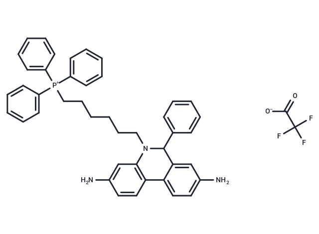
| Description | MitoSOX Red TFA (Mito-hydroethidine TFA) is a fluorescent probe that specifically targets mitochondria in living cells.MitoSOX Red is susceptible to oxidation by superoxide, but is not easily oxidized by other ROS or RNS-generating systems.MitoSOX Red acts as a fluorescent indicator and can be used for live cell imaging. |
| Synonyms | Mito-hydroethidine TFA(1003197-00-9 Free base), Mito-hydroethidine TFA |
| Cell Research | Instructions I. Solution preparation 1. Stock solution: MitoSOX Red TFA is usually dissolved in DMSO (dimethyl sulfoxide) to make a stock solution with a common concentration of 1 mM. When storing the stock solution, it should be stored at -20°C away from light and avoid repeated freezing and thawing to maintain stability. 2. Working solution: Dilute the stock solution to the required concentration, generally in the range of 1–5 µM, using an appropriate buffer such as PBS or culture medium. It is recommended to prepare the working solution freshly to ensure the best results. II. Cell staining steps 1. Cell preparation: For adherent cells, culture the cells on appropriate glass slides or culture plates. For suspended cells, use a suitable culture container. Make sure the cells are healthy and in the exponential growth phase for the best staining effect. 2. Staining steps: 1) Add the MitoSOX Red working solution directly to the cell culture medium, and the staining time is usually 10–30 minutes. The staining time can be optimized according to the cell type and experimental requirements. 2) During the staining process, avoid light exposure to prevent attenuation of the fluorescence signal. 3) Washing: After staining, remove the dye and wash the cells with PBS or appropriate culture medium to remove unbound dye. 4) Fluorescence microscopy: Observe cells using a fluorescence microscope, with an excitation wavelength of usually 510 nm and an emission wavelength of about 580 nm (red fluorescence), which can be used to observe superoxide in mitochondria. 5) Mitochondrial superoxide detection MitoSOX Red can be used to assess mitochondrial oxidative stress and superoxide production; the fluorescence intensity is related to the level of superoxide in mitochondria, so it can be used to study mitochondrial dysfunction, oxidative stress and related pathological processes. Precautions 1. Photosensitivity: MitoSOX Red is sensitive to light and should be avoided from prolonged exposure to strong light and stored in a light-proof environment to prevent attenuation of the fluorescence signal. 2. Toxicity: Use appropriate concentrations to avoid cytotoxicity. MitoSOX Red is generally safe at recommended concentrations, but excessive concentrations may cause cell damage, especially in long-term experiments. 3. Storage: MitoSOX Red stock solution should be stored at -20°C in the dark. Avoid repeated freezing and thawing as this may affect the stability and performance of the probe. 4. Mitochondrial membrane potential: Changes in mitochondrial membrane potential can affect the uptake of MitoSOX Red. Therefore, normal mitochondrial membrane potential should be ensured before the experiment to ensure effective staining. The above information is based on published literature. Experimental procedures should be appropriately modified to meet specific research demands. |
| Molecular Weight | 745.83 |
| Formula | C45H43F3N3O2P |
| Smiles | NC1=CC=C2C(C(C3=CC=CC=C3)N(CCCCCC[P+](C4=CC=CC=C4)(C5=CC=CC=C5)C6=CC=CC=C6)C7=C2C=CC(N)=C7)=C1.O=C(C(F)(F)F)[O-] |
| Relative Density. | no data available |
| Storage | Keep away from direct sunlight,Keep away from moisture,Store at low temperature,Store under nitrogen Store at -20°C Shipping with blue ice/Shipping at ambient temperature. |
 For example, if the intended dosage is 10 mg/kg for animals weighing 20 g , with a dosing volume of 100 μL per animal, and a total of 10 animals are to be administered, using a formulation of
  10% DMSO+ 40% PEG300+ 5% Tween 80+ 45% Saline/PBS/ddH2O , the resulting working solution concentration would be 2 mg/mL.
Dissolve 2 mg of the compound in 100 μL DMSO to obtain a stock solution at a concentration of 20 mg/mL . If the required concentration exceeds the compound's known solubility, please contact us for technical support before proceeding.
1) Add 100 μL of the DMSO stock solution to 400 µL PEG300 and mix thoroughly until the solution becomes clear.
2) Add 50 µL Tween 80 and mix well until fully clarified.
3) Add 450 µL Saline,PBS or ddH2O and mix thoroughly until a homogeneous solution is obtained.
| Size | Quantity | Unit Price | Amount | Operation |
|---|
 Hello! How can I help you today?
 Copyright © 2015-2026 TargetMol Chemicals Inc. All Rights Reserved.
