
Shopping Cart
Remove All Your shopping cart is currently empty

Synonyms: Vroac, Vrl2, Vanilloid receptor-related osmotically-activated channel (VR-OAC), Vanilloid receptor-like protein 2, Vanilloid receptor-like channel 2, Trpv4, Trp12, Transient receptor potential protein 12 (TRP12), Transient receptor potential cation channel subfamily V member 4, Osm-9-like TRP channel 4 (OTRPC4)
Anti-TRPV4 Polyclonal Antibody
| Pack Size | Price | USA Stock | Global Stock | Quantity |
|---|---|---|---|---|
| 50 µL | $221 | 7-10 days | 7-10 days | |
| 100 µL | $374 | 7-10 days | 7-10 days | |
| 200 µL | $527 | 7-10 days | 7-10 days |
| Description | Anti-TRPV4 Polyclonal Antibody is a Rabbit antibody targeting TRPV4. Anti-TRPV4 Polyclonal Antibody can be used in FCM, ICC/IF, IF, IHC-Fr, IHC-P. |
| Synonyms | Vroac, Vrl2, Vanilloid receptor-related osmotically-activated channel (VR-OAC), Vanilloid receptor-like protein 2, Vanilloid receptor-like channel 2, Trpv4, Trp12, Transient receptor potential protein 12 (TRP12), Transient receptor potential cation channel subfamily V member 4, Osm-9-like TRP channel 4 (OTRPC4) |
| Ig Type | IgG |
| Reactivity | Human (predicted:Mouse,Rat,Chicken,Dog,Pig,Cow) |
| Verified Activity | 1. Black line: Positive blank control (Raji); Negative blank control (HL60) Green line: Primary Antibody (Rabbit Anti-TRP12 antibody (TMAB-01908)) Orange line: Isotype Control Antibody (Rabbit IgG). Blue line: Secondary Antibody (Goat anti-rabbit IgG-PE)/(Goat anti-rabbit IgG-AF647) Raji (Positive) and HL60 (Negative control) cells (black) were fixed with 4% PFA for 10 min at room temperature, permeabilized with PBST for 20 min at room temperature, and incubated in 5% BSA blocking buffer for 30 min at room temperature. Cells were then stained with TRP12 Antibody (TMAB-01908) at 1:50 dilution in blocking buffer and incubated for 30 min at room temperature, washed twice with 2% BSA in PBS, followed by secondary antibody (blue) incubation for 40 min at room temperature. Acquisitions of 20,000 events were performed. Cells stained with primary antibody (green), and isotype control (orange). 2. Blank control: Raji. Primary Antibody (green line): Rabbit Anti-TRP12 antibody (TMAB-01908) Dilution: 1 μg/10^6 cells; Isotype Control Antibody (orange line): Rabbit IgG. Secondary Antibody: Goat anti-rabbit IgG-PE Dilution: 1 μg/test. Protocol The cells were fixed with 4% PFA (10 min at room temperature) and then permeabilized with PBST for 20 min at room temperature. The cells were then incubated in 5% BSA to block non-specific protein-protein interactions for 30 min at at room temperature. Cells stained with Primary Antibody for 30 min at room temperature. The secondary antibody used for 40 min at room temperature. 3. Paraformaldehyde-fixed, paraffin embedded (Human brain glioma); Antigen retrieval by microwave in sodium citrate buffer (pH6.0); Block endogenous peroxidase by 3% hydrogen peroxide for 30 minutes; Blocking buffer (3% BSA) at RT for 30 min; Antibody incubation with (TRP12) Polyclonal Antibody, Unconjugated (TMAB-01908) at 1:400 overnight at 4°C, followed by conjugation to the secondary antibody (labeled with HRP) and DAB staining. 4. NIH/3T3 cell; 4% Paraformaldehyde-fixed; Triton X-100 at room temperature for 20 min; Blocking buffer (normal goat serum) at 37°C for 20 min; Antibody incubation with (TRP12/TRPV4) polyclonal Antibody, Unconjugated (TMAB-01908) 1:100, 90 minutes at 37°C; followed by a conjugated Goat Anti-Rabbit IgG antibody at 37°C for 90 minutes, DAPI (blue) was used to stain the cell nucleus. 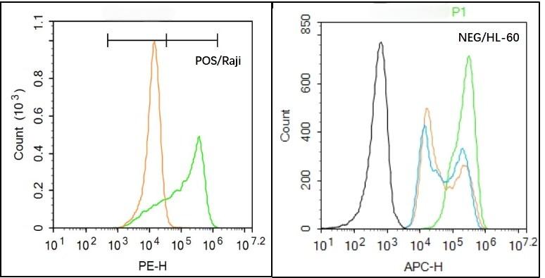 , ,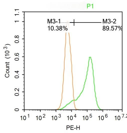 , ,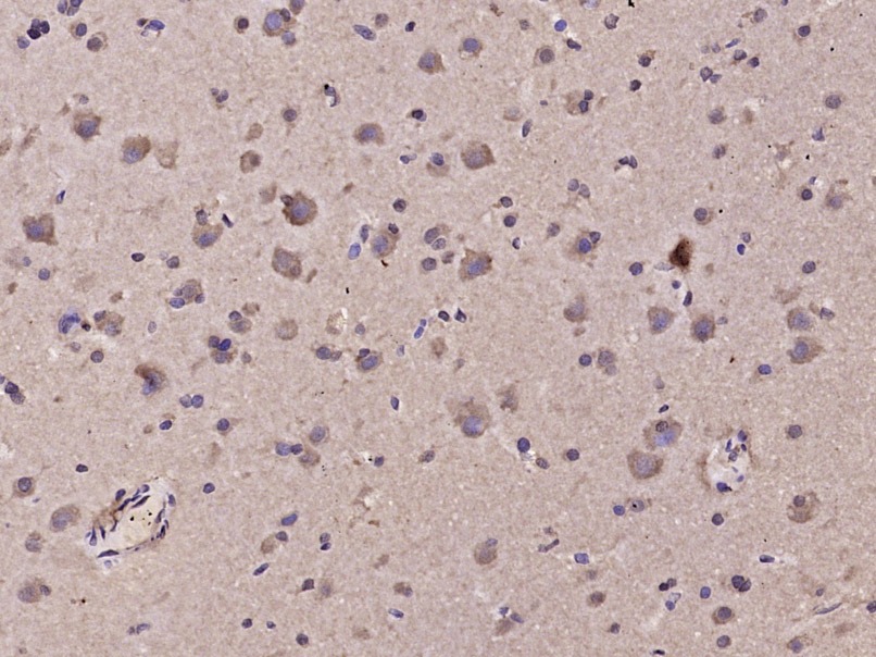 , ,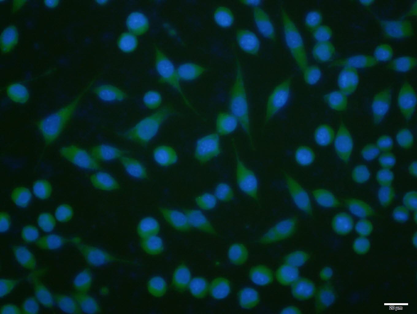 |
| Application | |
| Recommended Dose | FCM=1 μg/Test; ICC/IF=1:100-500; IF=1:100-500; IHC-Fr=1:100-500; IHC-P=1:100-500 |
| Antibody Type | Polyclonal |
| Host Species | Rabbit |
| Subcellular Localization | Cell membrane; Multi-pass membrane protein. Cell junction, adherens junction. Note=Assembly of the putative homotetramer occurs primarily in the endoplasmic reticulum. Isoform 1: Cell membrane. Isoform 5: Cell membrane. |
| Tissue Specificity | Found in the synoviocytes from patients with (RA) and without (CTR) rheumatoid arthritis (at protein level). |
| Construction | Polyclonal Antibody |
| Purification | Protein A purified |
| Appearance | Liquid |
| Formulation | 0.01M TBS (pH7.4) with 1% BSA, 0.02% Proclin300 and 50% Glycerol. |
| Concentration | 1 mg/mL |
| Research Background | The detection of noxious stimuli (chemical, mechanical, or thermal) occurs predominantly at the peripheral terminals of primary afferent neurons. This information is ultimately transmitted to the central nervous system to evoke appropriate protective reflexes. TRPV4 is a non selective calcium permeant, swell activated, cation channel probably involved in osmotic and mechano sensitivity. Activation by exposure to hypotonicity within the physiological range, low pH, citrate and phorbol esters exhibits an outward rectification. Once activated the channel seems to be regulated in a calmodulin dependent manner, with a negative feedback mechanism. |
| Immunogen | KLH conjugated synthetic peptide: human TRPV4 |
| Antigen Species | Human |
| Gene Name | TRPV4 |
| Gene ID | |
| Protein Name | Transient receptor potential cation channel subfamily V member 4 |
| Uniprot ID | |
| Biology Area | More Ion Channels,Nociception |
| Function | Non-selective calcium permeant cation channel probably involved in osmotic sensitivity and mechanosensitivity. Activation by exposure to hypotonicity within the physiological range exhibits an outward rectification. Also activated by low pH, citrate and phorbol esters. Increase of intracellular Ca(2+) potentiates currents. Channel activity seems to be regulated by a calmodulin-dependent mechanism with a negative feedback mechanism. Promotes cell-cell junction formation in skin keratinocytes and plays an important role in the formation and/or maintenance of functional intercellular barriers. Acts as a regulator of intracellular Ca(2+) in synoviocytes. Plays an obligatory role as a molecular component in the nonselective cation channel activation induced by 4-alpha-phorbol 12,13-didecanoate and hypotonic stimulation in synoviocytes and also regulates production of IL-8. |
| Molecular Weight | Theoretical: 96 kDa. |
| Stability & Storage | Store at -20°C or -80°C for 12 months. Avoid repeated freeze-thaw cycles. |
| Transport | Shipping with blue ice. |
| Size | Quantity | Unit Price | Amount | Operation |
|---|
 Hello! How can I help you today?
 Copyright © 2015-2026 TargetMol Chemicals Inc. All Rights Reserved.
