
Shopping Cart
Remove All Your shopping cart is currently empty

Synonyms: Transforming growth factor beta-1 proprotein
Anti-TGF beta 1 Antibody
(3J953)
| Pack Size | Price | USA Stock | Global Stock | Quantity |
|---|---|---|---|---|
| 50 µL | $298 | 7-10 days | 7-10 days | |
| 100 µL | $497 | 7-10 days | 7-10 days |
| Description | Anti-TGF beta 1 Antibody (3J953) is a Rabbit antibody targeting TGF beta 1. Anti-TGF beta 1 Antibody (3J953) can be used in IF-Tissue,IHC-P,WB. |
| Synonyms | Transforming growth factor beta-1 proprotein |
| Ig Type | IgG |
| Clone | 3J953 |
| Reactivity | Human,Mouse,Rat |
| Verified Activity | 1. Western blot analysis of TGF beta 1 on different lysates with Rabbit anti-TGF beta 1 antibody at 1/2,000 dilution. Lane 1: Raji cell lysate (15 μg/Lane) Lane 2: K-562 cell lysate (15 μg/Lane) Lane 3: Mouse spleen tissue lysate (20 μg/Lane) Lane 4: Rat spleen tissue lysate (20 μg/Lane) Predicted band size: 44 kDa Observed band size: 44 kDa Exposure time: 3 minutes 54 seconds; 4-20% SDS-PAGE gel. 2. Immunohistochemical analysis of paraffin-embedded rat spleen tissue with Rabbit anti-TGF beta 1 antibody at 1/200 dilution. The section was pre-treated using heat mediated antigen retrieval with Tris-EDTA buffer (pH 9.0) for 20 minutes. The tissues were blocked in 1% BSA for 20 minutes at room temperature, washed with ddH2O and PBS, and then probed with the primary antibody at 1/200 dilution for 1 hour at room temperature. The detection was performed using an HRP conjugated compact polymer system. DAB was used as the chromogen. Tissues were counterstained with hematoxylin and mounted with DPX. 3. Immunohistochemical analysis of paraffin-embedded mouse spleen tissue with Rabbit anti-TGF beta 1 antibody at 1/200 dilution. The section was pre-treated using heat mediated antigen retrieval with Tris-EDTA buffer (pH 9.0) for 20 minutes. The tissues were blocked in 1% BSA for 20 minutes at room temperature, washed with ddH2O and PBS, and then probed with the primary antibody at 1/200 dilution for 1 hour at room temperature. The detection was performed using an HRP conjugated compact polymer system. DAB was used as the chromogen. Tissues were counterstained with hematoxylin and mounted with DPX. 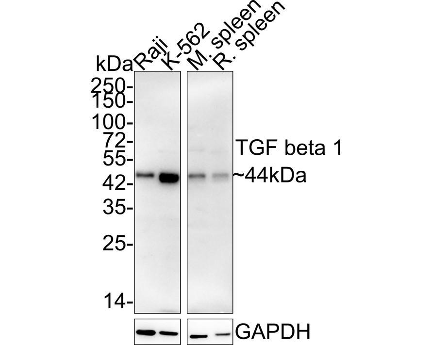 , ,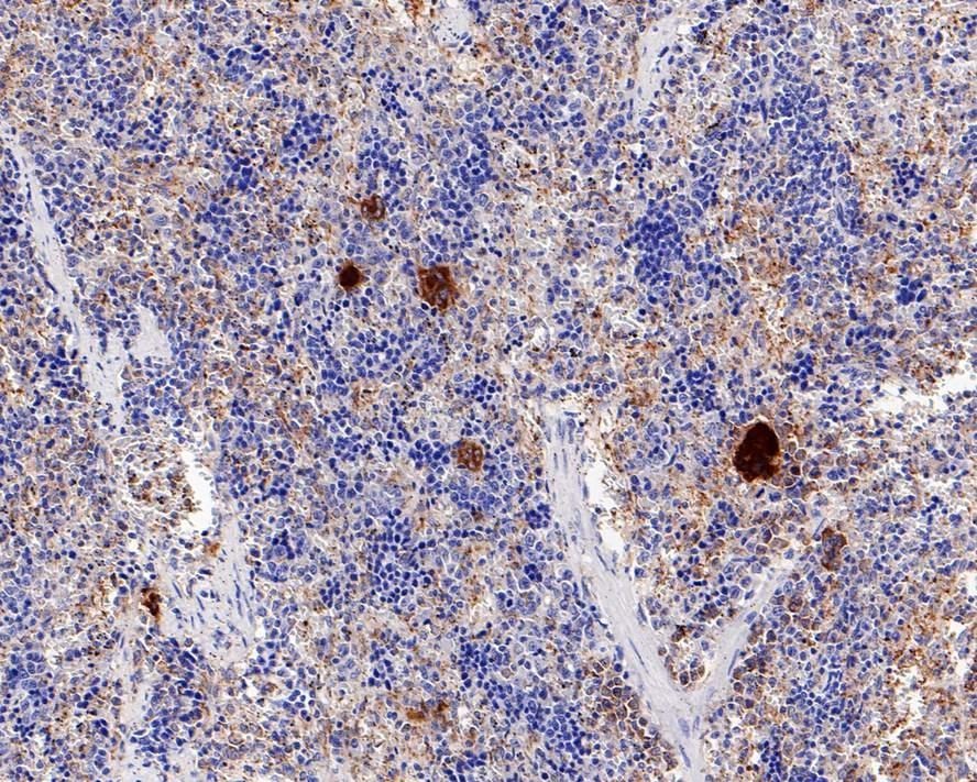 , ,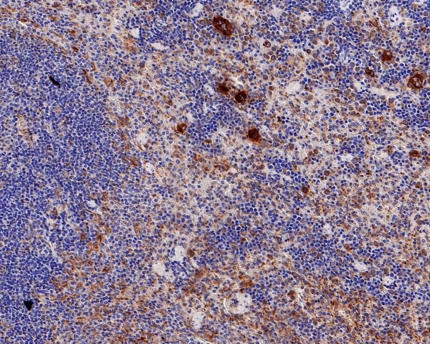 |
| Application | |
| Recommended Dose | WB: 1:2000; IHC-P: 1:200; IF-Tissue: 1:200 |
| Antibody Type | Monoclonal |
| Host Species | Rabbit |
| Construction | Recombinant Antibody |
| Purification | Protein A affinity purified. |
| Appearance | Liquid |
| Formulation | PBS (pH7.4), 0.1% BSA, 40% Glycerol. Preservative: 0.05% Sodium Azide. |
| Research Background | Many cells synthesize TGFB1 and have specific receptors for it. It positively and negatively regulates many other growth factors. It plays an important role in bone remodeling as it is a potent stimulator of osteoblastic bone formation, causing chemotaxis, proliferation and differentiation in committed osteoblasts. Heterodimers of TGFB1/TGFB2 have been found in bone. Interacts with CD109 and DPT. Interacts with ASPN. Function: TGF beta 2 has suppressive effects on interleukin-2 dependent T-cell growth. Tissue specificity: TGF beta 1 is highly expressed in bone. A chromosomal aberration involving TGFB2 is found in a family with Peters anomaly. |
| Conjucates | Unconjugated |
| Immunogen | KLH conjugated synthetic peptide: human TGF-Beta 1 |
| Antigen Species | Human |
| Uniprot ID |
| Molecular Weight | Theoretical: 44 kDa. |
| Stability & Storage | Store at 2°C-8°C for 1 month. Store at -20°C or -80°C for 12 months. Avoid repeated freeze-thaw cycles. |
| Transport | Shipping with blue ice. |
| Size | Quantity | Unit Price | Amount | Operation |
|---|
 Hello! How can I help you today?
 Copyright © 2015-2026 TargetMol Chemicals Inc. All Rights Reserved.
