Shopping Cart
Remove All Your shopping cart is currently empty
Anti-SDHA Antibody (3R603) is a Rabbit antibody targeting SDHA. Anti-SDHA Antibody (3R603) can be used in FCM,ICC/IF,IHC,WB.
| Pack Size | Price | USA Warehouse | Global Warehouse | Quantity |
|---|---|---|---|---|
| 50 μL | $298 | 7-10 days | 7-10 days | |
| 100 μL | $496 | 7-10 days | 7-10 days |
| Description | Anti-SDHA Antibody (3R603) is a Rabbit antibody targeting SDHA. Anti-SDHA Antibody (3R603) can be used in FCM,ICC/IF,IHC,WB. |
| Synonyms | Succinate dehydrogenase [ubiquinone] flavoprotein subunit, mitochondrial, SDHF, SDHA, SDH2, mitochondrial, Malate dehydrogenase [quinone] flavoprotein subunit, Flavoprotein subunit of complex II (Fp) |
| Ig Type | IgG |
| Clone | 3R603 |
| Reactivity | Human,Mouse,Rat,zebrafish |
| Verified Activity | 1. Western blot analysis of SDHA on Jurkat cells lysates using anti-SDHA antibody at 1/500 dilution. 2. Immunohistochemical analysis of paraffin-embedded zebrafish tissue using anti-SDHA antibody. Counter stained with hematoxylin. 3. Immunohistochemical analysis of paraffin-embedded human liver tissue using anti-SDHA antibody. Counter stained with hematoxylin. 4. Immunohistochemical analysis of paraffin-embedded human kidney tissue using anti-SDHA antibody. Counter stained with hematoxylin. 5. Immunohistochemical analysis of paraffin-embedded mouse testis tissue using anti-SDHA antibody. Counter stained with hematoxylin. 6. Immunohistochemical analysis of paraffin-embedded mouse skeletal muscle tissue using anti-SDHA antibody. Counter stained with hematoxylin. 7. Immunohistochemical analysis of paraffin-embedded mouse colon tissue using anti-SDHA antibody. Counter stained with hematoxylin. 8. ICC staining SDHA in MCF-7 cells (red). The nuclear counter stain is DAPI (blue). Cells were fixed in paraformaldehyde, permeabilised with 0.25% Triton X100/PBS. 9. ICC staining SDHA in HepG2 cells (red). The nuclear counter stain is DAPI (blue). Cells were fixed in paraformaldehyde, permeabilised with 0.25% Triton X100/PBS. 10. ICC staining SDHA in NIH-3T3 cells (red). The nuclear counter stain is DAPI (blue). Cells were fixed in paraformaldehyde, permeabilised with 0.25% Triton X100/PBS. 11. Flow cytometric analysis of Hela cells with SDHA antibody at 1/50 dilution (red) compared with an unlabelled control (cells without incubation with primary antibody; black). Alexa Fluor 488-conjugated goat anti rabbit IgG was used as the secondary antibody.  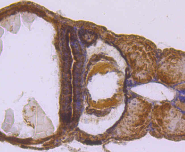 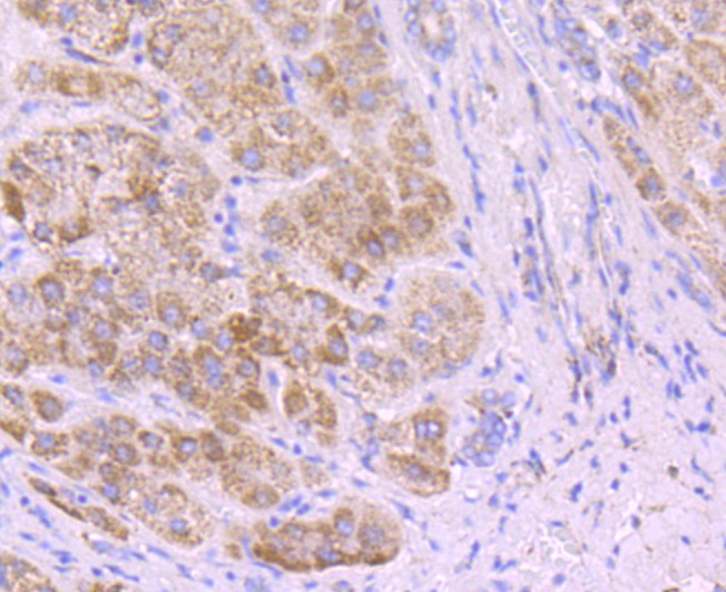 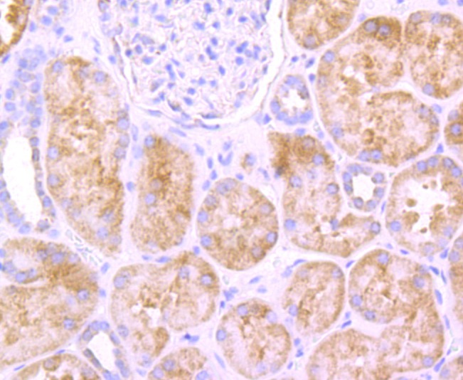 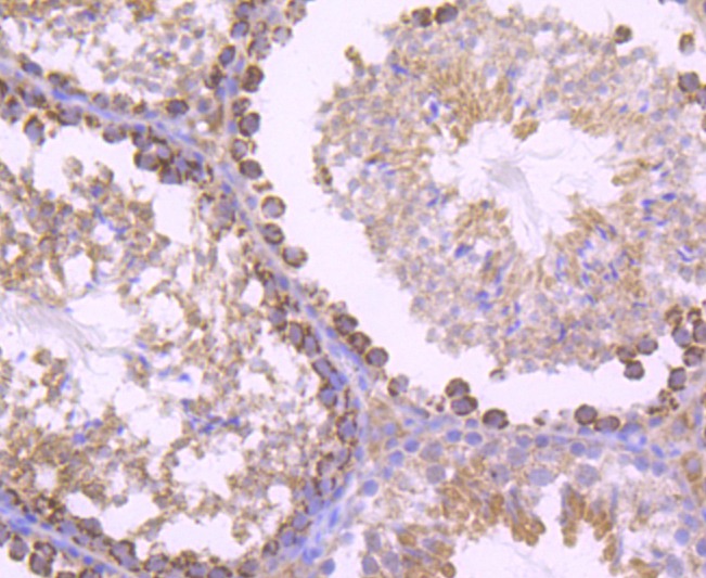 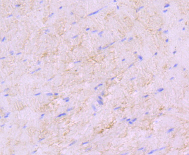 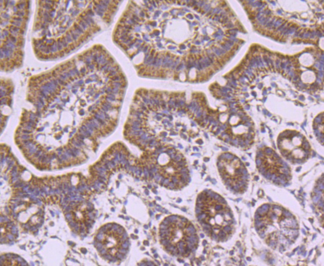 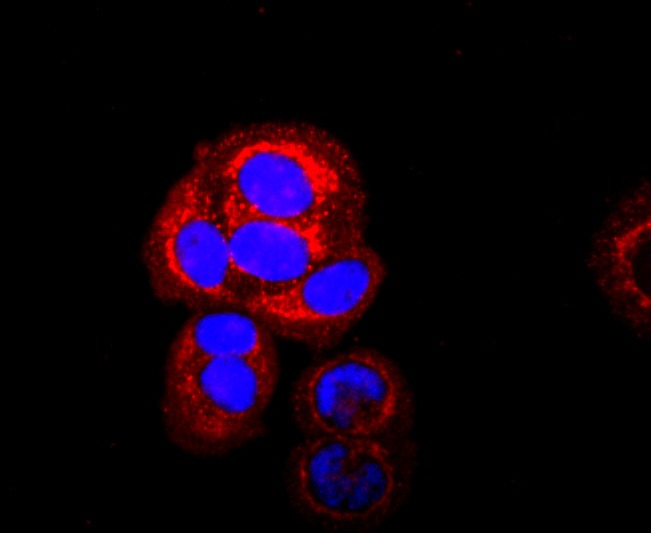 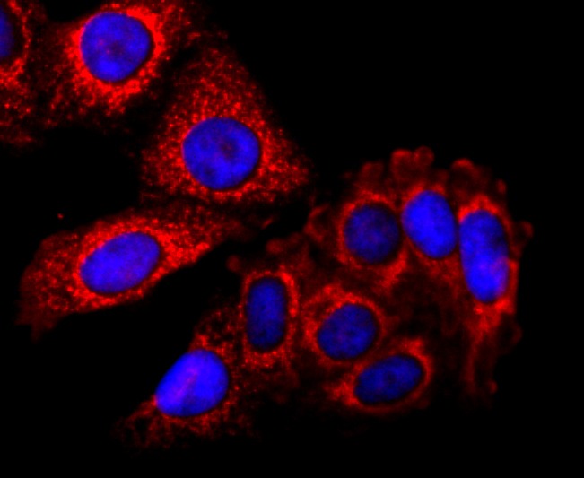 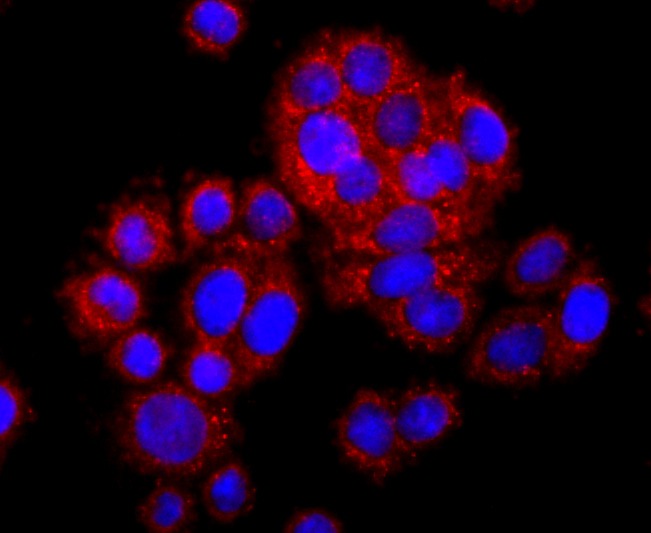 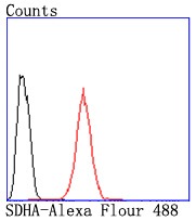 |
| Application | |
| Recommended Dose | WB: 1:500-1000; IHC: 1:50-200; ICC/IF: 1:50-200; FCM: 1:50-100 |
| Antibody Type | Monoclonal |
| Host Species | Rabbit |
| Construction | Recombinant Antibody |
| Purification | ProA affinity purified |
| Appearance | Liquid |
| Formulation | 1*TBS (pH7.4), 1%BSA, 40%Glycerol. Preservative: 0.05% Sodium Azide. |
| Research Background | In aerobic respiration reactions, succinate dehydrogenase (SDH) catalyzes the oxidation of succinate and ubiquinone to fumarate and ubiquinol. Four subunits comprise the SDH protein complex: a flavochrome subunit (SDHA), an iron-sulfur protein (SDHB), and two membrane-bound subunits (SDHC and SDHD) anchored to the inner mitochondrial membrane. Mutations to these subunits cause mitochondrial dysfunction, corresponding to several distinct disorders. Mutations in the membrane bound components may cause hereditary paraganglioma, while SDHA mutations are associated with juvenile encephalopathy as well as Leigh Syndrome, a severe neurological disorder. Inactivating mutations in SDHB correlate with inherited, but not necessarily sporadic, cases of pheochromocytoma. |
| Conjucates | Unconjugated |
| Immunogen | Recombinant Protein |
| Uniprot ID |
| Molecular Weight | Theoretical: 68 kDa. |
| Stability & Storage | Store at -20°C or -80°C for 12 months. Avoid repeated freeze-thaw cycles. |
| Transport | Shipping with blue ice. |
| Size | Quantity | Unit Price | Amount | Operation |
|---|

Copyright © 2015-2026 TargetMol Chemicals Inc. All Rights Reserved.