
Shopping Cart
Remove All Your shopping cart is currently empty

Synonyms: Related adhesion focal tyrosine kinase, RAFTK2, RAFTK, PYK 2, PTK2B protein tyrosine kinase 2 beta, PTK2B, PTK, Protein-tyrosine kinase 2-beta, Protein Tyrosine Kinase 2 Beta, Protein kinase B, Proline-rich tyrosine kinase 2, PKB, MGC124628, Focal adhesion kinase 2, FAK2_HUMAN, FAK2, FADK2, FADK 2, EC 2.7.10.2, E430023O05Rik, Cell adhesion kinase beta, Calcium-dependent tyrosine kinase, Calcium regulated non receptor proline rich tyrosine kinase, CAK-beta, CAKbeta, CAKB, CADTK
Anti-PYK2 Antibody
(5I30)
| Pack Size | Price | USA Stock | Global Stock | Quantity |
|---|---|---|---|---|
| 50 µL | $296 | 7-10 days | 7-10 days | |
| 100 µL | $498 | 7-10 days | 7-10 days |
| Description | Anti-PYK2 Antibody (5I30) is a Rabbit antibody targeting PYK2. Anti-PYK2 Antibody (5I30) can be used in ICC/IF,IHC,WB. |
| Synonyms | Related adhesion focal tyrosine kinase, RAFTK2, RAFTK, PYK 2, PTK2B protein tyrosine kinase 2 beta, PTK2B, PTK, Protein-tyrosine kinase 2-beta, Protein Tyrosine Kinase 2 Beta, Protein kinase B, Proline-rich tyrosine kinase 2, PKB, MGC124628, Focal adhesion kinase 2, FAK2_HUMAN, FAK2, FADK2, FADK 2, EC 2.7.10.2, E430023O05Rik, Cell adhesion kinase beta, Calcium-dependent tyrosine kinase, Calcium regulated non receptor proline rich tyrosine kinase, CAK-beta, CAKbeta, CAKB, CADTK |
| Ig Type | IgG |
| Clone | 5I30 |
| Reactivity | Human,Mouse,Rat |
| Verified Activity | 1. Western blot analysis of PYK2 on mouse brain lysates using anti-PYK2 antibody at 1/1,000 dilution. 2. Immunohistochemical analysis of paraffin-embedded human tonsil tissue using anti-PYK2 antibody. Counter stained with hematoxylin. 3. Immunohistochemical analysis of paraffin-embedded human spleen tissue using anti-PYK2 antibody. Counter stained with hematoxylin. 4. Immunohistochemical analysis of paraffin-embedded human kidney tissue using anti-PYK2 antibody. Counter stained with hematoxylin. 5. Immunohistochemical analysis of paraffin-embedded mouse brain tissue using anti-PYK2 antibody. Counter stained with hematoxylin. 6. Immunohistochemical analysis of paraffin-embedded mouse kidney tissue using anti-PYK2 antibody. Counter stained with hematoxylin. 7. ICC staining PYK2 in Hela cells (green). The nuclear counter stain is DAPI (blue). Cells were fixed in paraformaldehyde, permeabilised with 0.25% Triton X100/PBS. 8. ICC staining PYK2 in SHG-44 cells (green). The nuclear counter stain is DAPI (blue). Cells were fixed in paraformaldehyde, permeabilised with 0.25% Triton X100/PBS. 9. ICC staining PYK2 in SH-SY-5Y cells (green). The nuclear counter stain is DAPI (blue). Cells were fixed in paraformaldehyde, permeabilised with 0.25% Triton X100/PBS. 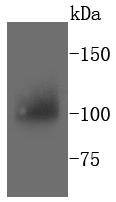 , ,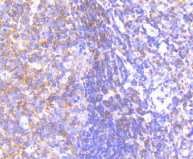 , ,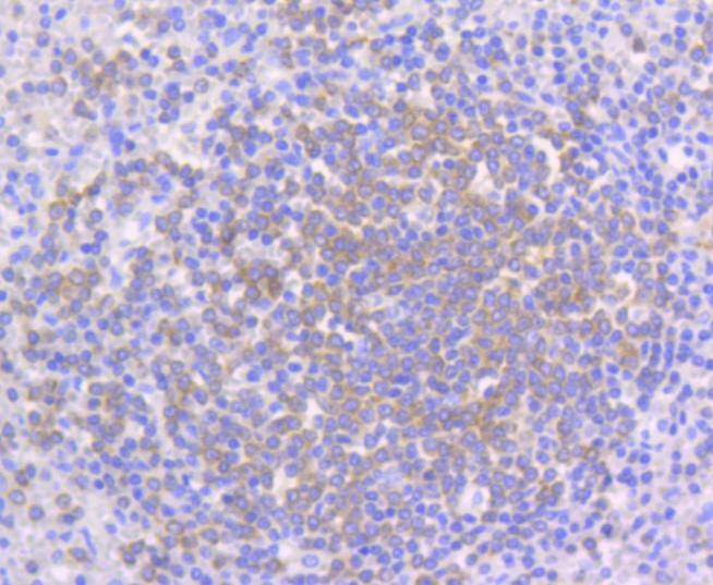 , ,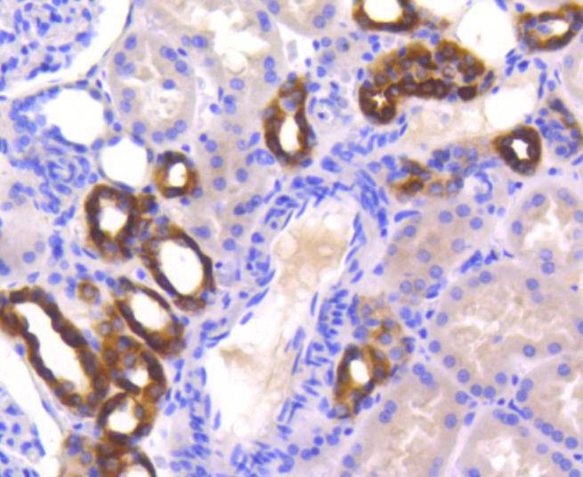 , ,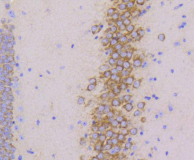 , ,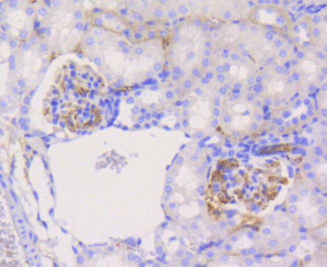 , ,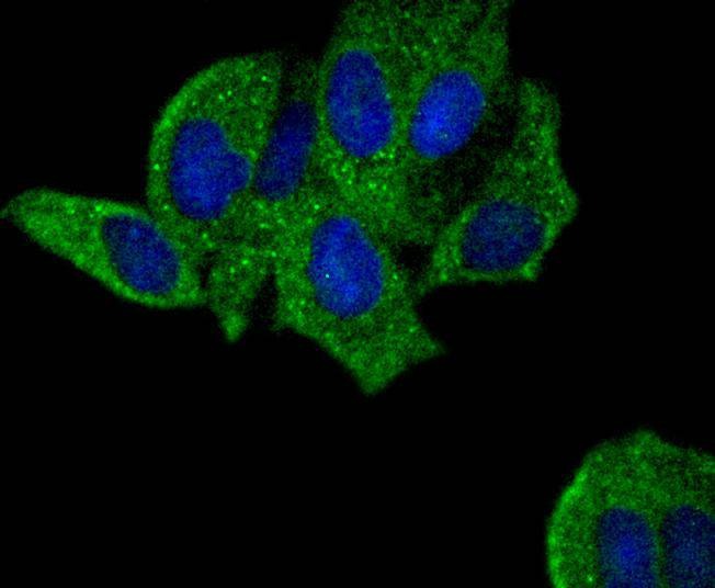 , ,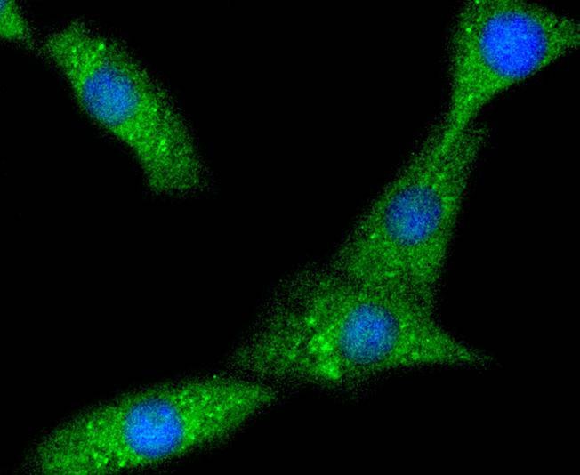 , ,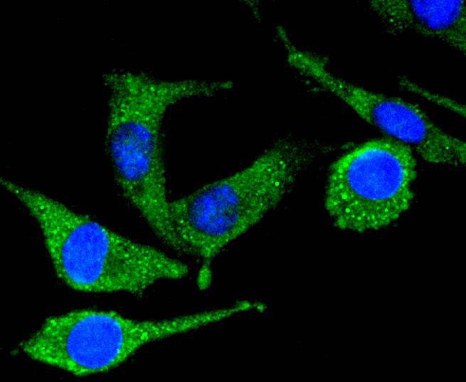 |
| Application | |
| Recommended Dose | WB: 1:1000-2000; IHC: 1:50-200; ICC/IF: 1:50-200 |
| Antibody Type | Monoclonal |
| Host Species | Rabbit |
| Construction | Recombinant Antibody |
| Purification | ProA affinity purified |
| Appearance | Liquid |
| Formulation | 1*TBS (pH7.4), 1%BSA, 40%Glycerol. Preservative: 0.05% Sodium Azide. |
| Research Background | Focal adhesion kinase (FAK) was initially identified as a substrate for the intrinsic protein tyrosine kinase activity of Src-encoded pp60. The deduced amino acid sequence of FAK p125 has shown it to be a cytoplasmic protein tyrosine kinase whose sequence and structural organization are unique compared to other protein families described. A putative new member of the FAK family, designated PYK2 (proline-rich tyrosine kinase 2), exhibits 61% sequence identity with FAK over its kinase domain. PYK2 (also designated CAKb or RAFTK) is highly expressed in the central nervous system. Activation of the kinase leads to modulation of ion channel function and the activation of the MAPK signaling pathway. PYK2 is rapidly phosphorylated on tyrosine residues in response to stimuli that increase intracellular calcium levels and compounds that activate members of the PKC family of kinases, such as phorbol esters. |
| Conjucates | Unconjugated |
| Immunogen | Recombinant Protein |
| Uniprot ID |
| Molecular Weight | Theoretical: 116 kDa. |
| Stability & Storage | Store at -20°C or -80°C for 12 months. Avoid repeated freeze-thaw cycles. |
| Transport | Shipping with blue ice. |
| Size | Quantity | Unit Price | Amount | Operation |
|---|
 Hello! How can I help you today?
 Copyright © 2015-2026 TargetMol Chemicals Inc. All Rights Reserved.
