
Shopping Cart
Remove All Your shopping cart is currently empty

Synonyms: p-IRF7 (Ser471, 472), p-IRF7 (S471, 472), IRF7 (p-Ser471, 472), IRF7 (p-S471, 472), IRF-7, IRF7, Interferon regulatory factor 7
Anti-Phospho-IRF7
(Ser471, 472) Polyclonal Antibody
| Pack Size | Price | USA Stock | Global Stock | Quantity |
|---|---|---|---|---|
| 50 µL | $220 | 7-10 days | 7-10 days | |
| 100 µL | $374 | 7-10 days | 7-10 days | |
| 200 µL | $527 | 7-10 days | 7-10 days |
| Description | Anti-Phospho-IRF7 (Ser471, 472) Polyclonal Antibody is a Rabbit antibody targeting Phospho-IRF7 (Ser471, 472). Anti-Phospho-IRF7 (Ser471, 472) Polyclonal Antibody can be used in ICC/IF, IF, IHC-Fr, IHC-P, WB. |
| Synonyms | p-IRF7 (Ser471, 472), p-IRF7 (S471, 472), IRF7 (p-Ser471, 472), IRF7 (p-S471, 472), IRF-7, IRF7, Interferon regulatory factor 7 |
| Ig Type | IgG |
| Reactivity | Human,Mouse,Rat (predicted:Pig,Cow,Horse) |
| Verified Activity | 1. HepG2 cell; 4% Paraformaldehyde-fixed; Triton X-100 at room temperature for 20 min; Blocking buffer (normal goat serum) at 37°C for 20 min; Antibody incubation with (Phospho-IRF7 (Ser471 + Ser472)) polyclonal Antibody, Unconjugated (TMAB-01437) 1:100, 90 minutes at 37°C; followed by a conjugated Goat Anti-Rabbit IgG antibody at 37°C for 90 minutes, DAPI (blue) was used to stain the cell nucleus. 2. Paraformaldehyde-fixed, paraffin embedded (Human brain glioma); Antigen retrieval by boiling in sodium citrate buffer (pH6.0) for 15 min; Block endogenous peroxidase by 3% hydrogen peroxide for 20 min; Blocking buffer (normal goat serum) at 37°C for 30 min; Antibody incubation with (Phospho-IRF7 (Ser471 + Ser472)) Polyclonal Antibody, Unconjugated (TMAB-01437) at 1:400 overnight at 4°C, followed by operating according to SP Kit (Rabbit) instructionsand DAB staining. 3. Paraformaldehyde-fixed, paraffin embedded (Mouse brain); Antigen retrieval by boiling in sodium citrate buffer (pH6.0) for 15 min; Block endogenous peroxidase by 3% hydrogen peroxide for 20 min; Blocking buffer (normal goat serum) at 37°C for 30 min; Antibody incubation with (Phospho-IRF7 (Ser471 + Ser472)) Polyclonal Antibody, Unconjugated (TMAB-01437) at 1:400 overnight at 4°C, followed by operating according to SP Kit (Rabbit) instructionsand DAB staining. 4. Sample: Lane 1: Mouse Spleen tissue lysates Lane 2: Mouse Thymus tissue lysates Lane 3: Mouse Blood cell lysates Lane 4: Mouse Lung tissue lysates Lane 5: Rat Spleen tissue lysates Lane 6: Rat Liver tissue lysates Lane 7: Human Jurkat cell lysates Lane 8: Human Raji cell lysates Primary: Anti-Phospho-IRF7 (Ser471 + Ser472) (TMAB-01437) at 1/1000 dilution Secondary: IRDye800CW Goat Anti-Rabbit IgG at 1/20000 dilution Predicted band size: 54 kDa Observed band size: 70,55 kDa 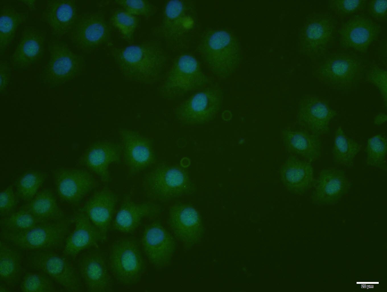 , ,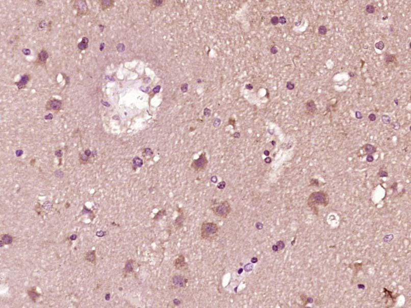 , ,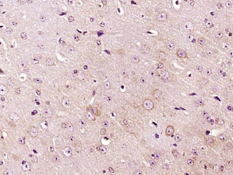 , ,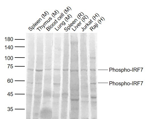 |
| Application | |
| Recommended Dose | ICC/IF=1:100-500; IF=1:100-500; IHC-Fr=1:100-500; IHC-P=1:100-500; WB=1:500-2000 |
| Antibody Type | Polyclonal |
| Host Species | Rabbit |
| Subcellular Localization | Nucleus. Cytoplasm. Note=The phosphorylated and active form accumulates selectively in the nucleus. |
| Tissue Specificity | Expressed predominantly in spleen, thymus and peripheral blood leukocytes. |
| Construction | Polyclonal Antibody |
| Purification | Protein A purified |
| Appearance | Liquid |
| Formulation | 0.01M TBS (pH7.4) with 1% BSA, 0.02% Proclin300 and 50% Glycerol. |
| Concentration | 1 mg/mL |
| Research Background | IRF7 encodes interferon regulatory factor 7, a member of the interferon regulatory transcription factor (IRF) family. IRF7 has been shown to play a role in the transcriptional activation of virus-inducible cellular genes, including interferon beta chain genes. Inducible expression of IRF7 is largely restricted to lymphoid tissue. Multiple IRF7 transcript variants have been identified, although the functional consequences of these have not yet been established. [provided by RefSeq, Jul 2008] |
| Immunogen | KLH conjugated synthesised phosphopeptide: human IRF7 around the phosphorylation site of Ser471/472 |
| Antigen Species | Human |
| Gene Name | IRF7 |
| Gene ID | |
| Protein Name | Interferon regulatory factor 7 |
| Uniprot ID | |
| Biology Area | Helix-Turn-Helix,Pol II Transcription,Antiviral Signaling,Interferons,TLR Signaling,Host Virus Interaction,SARS Coronavirus |
| Function | Key transcriptional regulator of type I interferon (IFN)-dependent immune responses and plays a critical role in the innate immune response against DNA and RNA viruses. Regulates the transcription of type I IFN genes (IFN-alpha and IFN-beta) and IFN-stimulated genes (ISG) by binding to an interferon-stimulated response element (ISRE) in their promoters. Can efficiently activate both the IFN-beta (IFNB) and the IFN-alpha (IFNA) genes and mediate their induction via both the virus-activated, MyD88-independent pathway and the TLR-activated, MyD88-dependent pathway. Required during both the early and late phases of the IFN gene induction but is more critical for the late than for the early phase. Exists in an inactive form in the cytoplasm of uninfected cells and following viral infection, double-stranded RNA (dsRNA), or toll-like receptor (TLR) signaling, becomes phosphorylated by IKBKE and TBK1 kinases. This induces a conformational change, leading to its dimerization and nuclear localization where along with other coactivators it can activate transcription of the type I IFN and ISG genes. Can also play a role in regulating adaptive immune responses by inducing PSMB9/LMP2 expression, either directly or through induction of IRF1. Binds to the Q promoter (Qp) of EBV nuclear antigen 1 a (EBNA1) and may play a role in the regulation of EBV latency. Can activate distinct gene expression programs in macrophages and regulate the anti-tumor properties of primary macrophages. |
| Molecular Weight | Theoretical: 54 kDa. Actual: 70,55 kDa. |
| Stability & Storage | Store at -20°C or -80°C for 12 months. Avoid repeated freeze-thaw cycles. |
| Transport | Shipping with blue ice. |
| Size | Quantity | Unit Price | Amount | Operation |
|---|
 Hello! How can I help you today?
 Copyright © 2015-2026 TargetMol Chemicals Inc. All Rights Reserved.
