
Shopping Cart
Remove All Your shopping cart is currently empty


Anti-p53 Polyclonal Antibody is a Rabbit antibody targeting p53. Anti-p53 Polyclonal Antibody can be used in FCM,ICC/IF,IF,IHC-Fr,IHC-P,WB.
| Pack Size | Price | USA Warehouse | Global Warehouse | Quantity |
|---|---|---|---|---|
| 50 μL | $220 | 7-10 days | 7-10 days | |
| 100 μL | $374 | 7-10 days | 7-10 days | |
| 200 μL | $528 | 7-10 days | 7-10 days |
| Description | Anti-p53 Polyclonal Antibody is a Rabbit antibody targeting p53. Anti-p53 Polyclonal Antibody can be used in FCM,ICC/IF,IF,IHC-Fr,IHC-P,WB. |
| Ig Type | IgG |
| Reactivity | Human,Mouse,Rat (predicted:Pig,Cow,Horse,Rabbit,Sheep) |
| Verified Activity | 1. Paraformaldehyde-fixed, paraffin embedded (Rat bladder); Antigen retrieval by boiling in sodium citrate buffer (pH6.0) for 15 min; Block endogenous peroxidase by 3% hydrogen peroxide for 20 min; Blocking buffer (normal goat serum) at 37°C for 30 min; Antibody incubation with (P53 protein (wt-p53)) Polyclonal Antibody, Unconjugated (TMAB-01319) at 1:200 overnight at 4°C, followed by operating according to SP Kit (Rabbit) instructions and DAB staining. 2. Paraformaldehyde-fixed, paraffin embedded (Rat esophageal); Antigen retrieval by boiling in sodium citrate buffer (pH6.0) for 15 min; Block endogenous peroxidase by 3% hydrogen peroxide for 20 min; Blocking buffer (normal goat serum) at 37°C for 30 min; Antibody incubation with (P53 protein (wt-p53)) Polyclonal Antibody, Unconjugated (TMAB-01319) at 1:200 overnight at 4°C, followed by operating according to SP Kit (Rabbit) instructions and DAB staining. 3. Paraformaldehyde-fixed, paraffin embedded (mouse esophageal); Antigen retrieval by boiling in sodium citrate buffer (pH6.0) for 15 min; Block endogenous peroxidase by 3% hydrogen peroxide for 20 min; Blocking buffer (normal goat serum) at 37°C for 30 min; Antibody incubation with (P53 protein (wt-p53)) Polyclonal Antibody, Unconjugated (TMAB-01319) at 1:200 overnight at 4°C, followed by operating according to SP Kit (Rabbit) instructions and DAB staining. 4. Sample: Liver (Mouse) Lysate at 40 μg Primary: Anti-P53 protein (wt-p53)(TMAB-01319) at 1/300 dilution Secondary: IRDye800CW Goat Anti-Rabbit IgG at 1/20000 dilution Predicted band size: 43 kDa Observed band size: 50 kDa 5. Sample: Brain (Rat) lysate at 30 μg; Colon carcinoma (Human) lysate at30 μg; Primary: Anti-wt-p53 (TMAB-01319) at 1:200 dilution; Secondary: HRP conjugated Goat-Anti-Rabbit Igg at 1: 3000 dilution; Predicted band size: 43 kDa Observed band size: 53 kDa 6. Sample: HepG2 Cell Lysate at 40 μg Mcf-7 Cell Lysate at 40 μg DU145 Cell Lysate at 40 μg Primary: Anti-P53 protein (wt-p53)(TMAB-01319) at 1/300 dilution Secondary: IRDye800CW Goat Anti-Rabbit IgG at 1/20000 dilution Predicted band size: 43 kDa Observed band size: 50 kDa 7. Independently Validated Antibody, image provided by Science Direct, badge number 029660. Formalin-fixed and paraffin embedded human testis and breast tissue stained with Rabbit Anti-P53 protein (wt-p53) Polyclonal Antibody at 1:250 at room temperature overnight. Both positive and negative controls stained. 8. Blank control (blue line): HeLa (blue). Primary Antibody (green line): Rabbit Anti-P53 protein (wt-p53) antibody (TMAB-01319), Dilution: 1 μg/10^6 cells; Isotype Control Antibody (orange line): Rabbit IgG. Secondary Antibody (white blue line): Goat anti-rabbit IgG-FITC Dilution: 1 μg/test. Protocol The cells were fixed with 70% ethanol (Overnight at 4°C) and then permeabilized with 90% ice-cold methanol for 30 min on ice. Cells stained with Primary Antibody for 30 min at room temperature. The cells were then incubated in 1 X PBS/2% BSA/10% goat serum to block non-specific protein-protein interactions followed by the antibody for 15 min at room temperature. The secondary antibody used for 40 min at room temperature. 9. Paraformaldehyde-fixed, paraffin embedded (Human breast cancer); Antigen retrieval by boiling in sodium citrate buffer (pH6.0) for 15 min; Block endogenous peroxidase by 3% hydrogen peroxide for 20 min; Blocking buffer (normal goat serum) at 37°C for 30 min; Antibody incubation with (P53 protein (wt-p53)) Polyclonal Antibody, Unconjugated (TMAB-01319) at 1:400 overnight at 4°C, followed by operating according to SP Kit (Rabbit) instructions and DAB staining. 10. Tissue/cell: A549 cell; 4% Paraformaldehyde-fixed; Triton X-100 at room temperature for 20 min; Blocking buffer (normal goat serum) at 37°C for 20 min; Antibody incubation with (P53 protein (wt-p53)) polyclonal Antibody, Unconjugated (TMAB-01319) 1:100, 90 minutes at 37°C; followed by a FITC conjugated Goat Anti-Rabbit IgG antibody at 37°C for 90 minutes, DAPI (blue) was used to stain the cell nucleus. 11. Tissue/cell: A549 cell; 4% Paraformaldehyde-fixed; Triton X-100 at room temperature for 20 min; Blocking buffer (normal goat serum) at 37°C for 20 min; Antibody incubation with (P53 protein (wt-p53)) polyclonal Antibody, Unconjugated (TMAB-01319) 1:100, 90 minutes at 37°C; followed by a FITC conjugated Goat Anti-Rabbit IgG antibody at 37°C for 90 minutes, DAPI (blue) was used to stain the cell nucleus. 12. Blank control: A549. Primary Antibody (green line): Rabbit Anti-P53 protein (wt-p53) antibody (TMAB-01319) Dilution: 1 μg/10^6 cells; Isotype Control Antibody (orange line): Rabbit IgG. Secondary Antibody: Goat anti-rabbit IgG-AF488 Dilution: 1 μg/test. Protocol The cells were fixed with 4% PFA (10 min at room temperature) and then permeabilized with 90% ice-cold methanol for 20 min at-20°C. The cells were then incubated in 5% BSA to block non-specific protein-protein interactions for 30 min at room temperature. Cells stained with Primary Antibody for 30 min at room temperature. The secondary antibody used for 40 min at room temperature. 13. Tissue/cell: A431 cell; 4% Paraformaldehyde-fixed; Triton X-100 at room temperature for 20 min; Blocking buffer (normal goat serum) at 37°C for 20 min; Antibody incubation with (P53 protein (wt-p53)) polyclonal Antibody, Unconjugated (TMAB-01319) 1:100, 90 minutes at 37°C; followed by a FITC conjugated Goat Anti-Rabbit IgG antibody at 37°C for 90 minutes, DAPI (blue) was used to stain the cell nucleus. 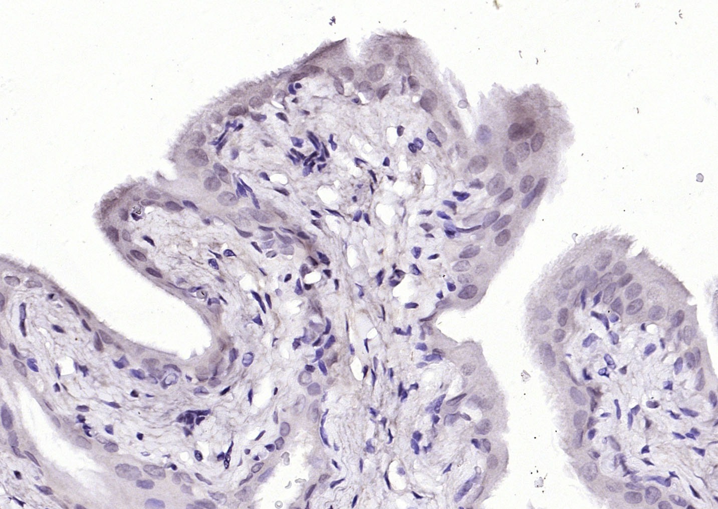 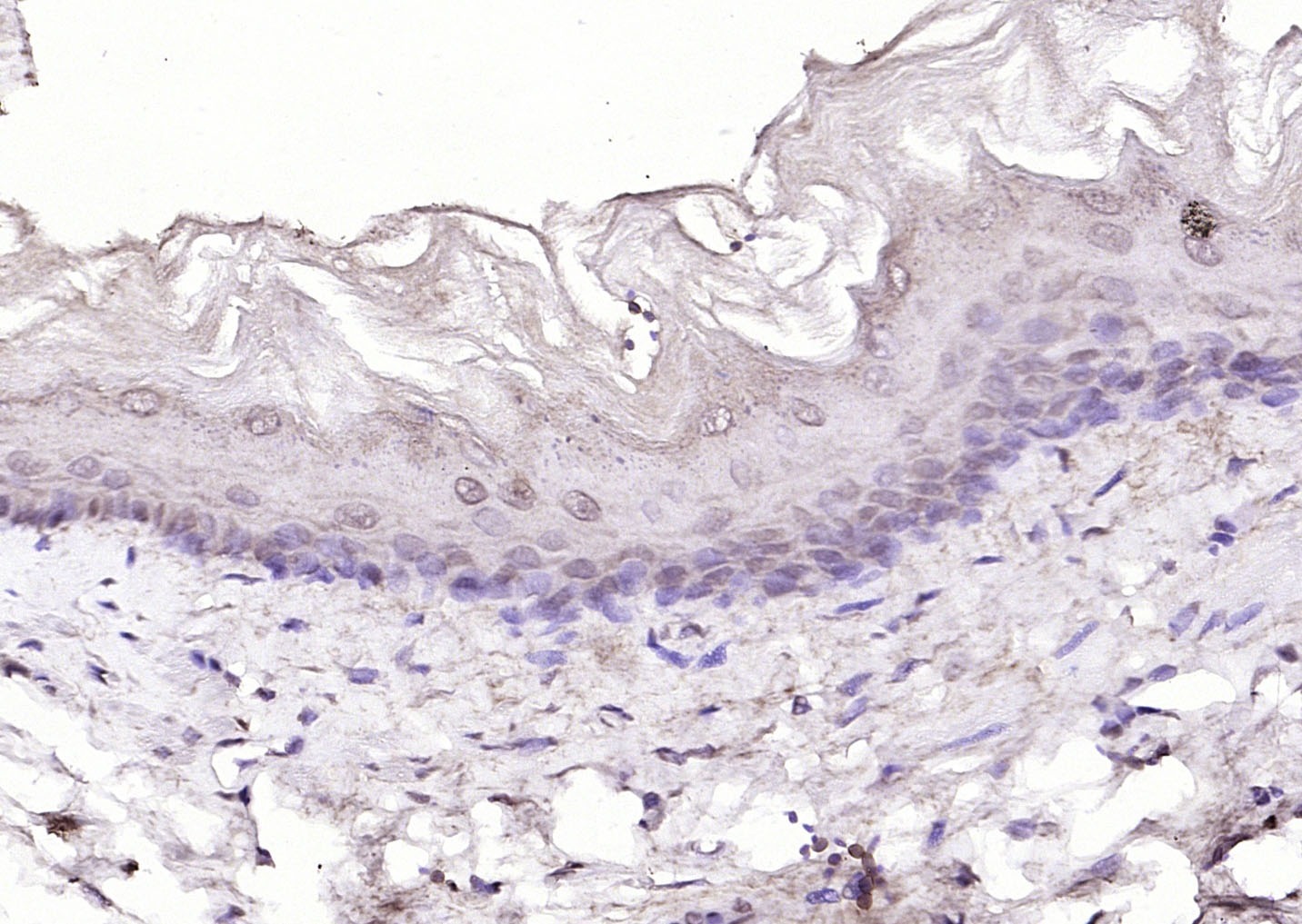 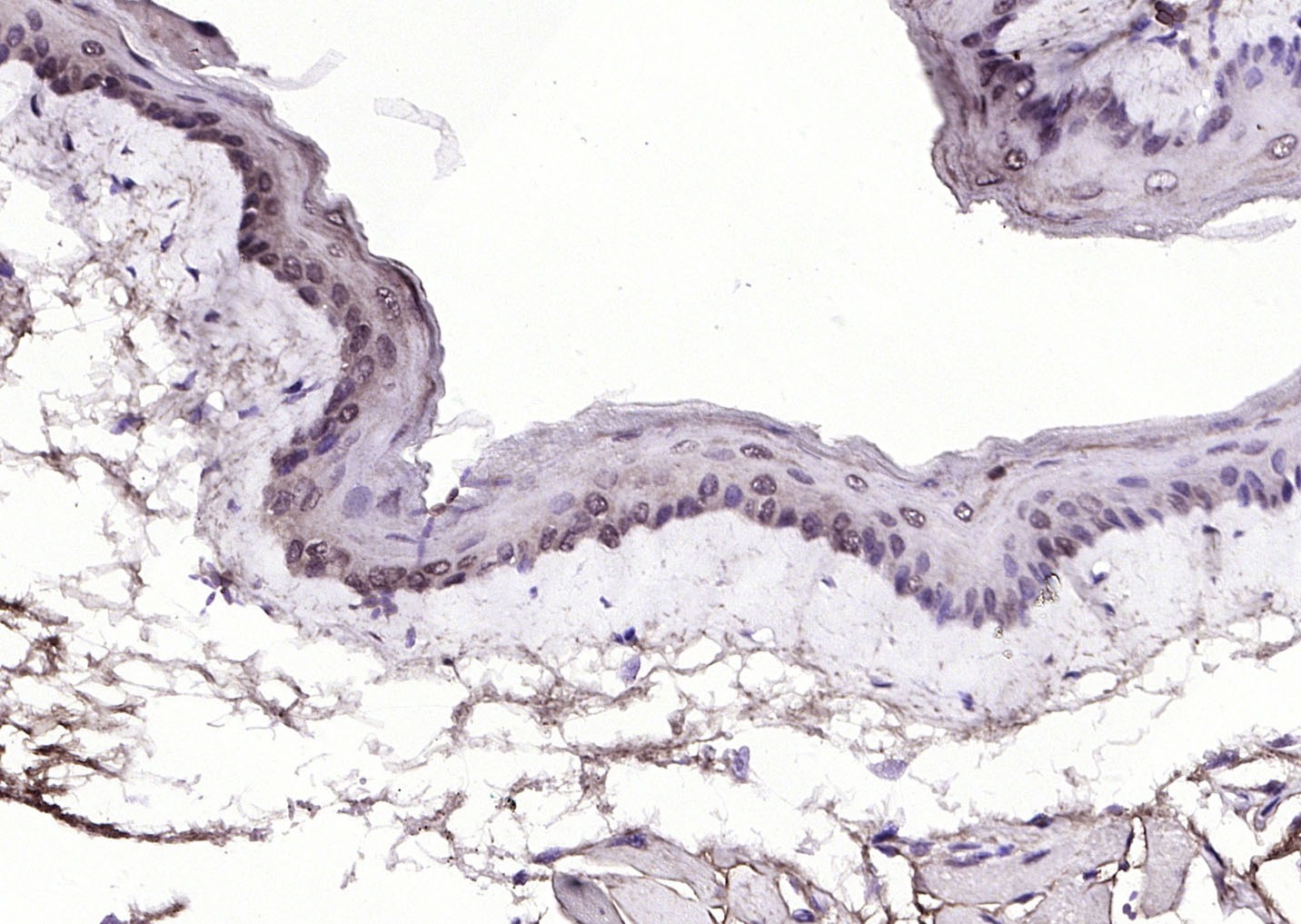 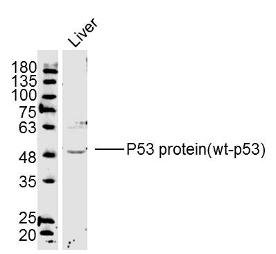 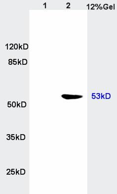 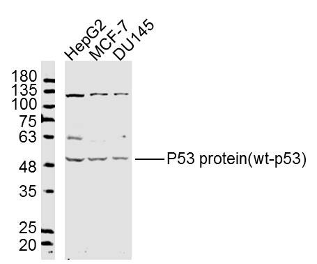 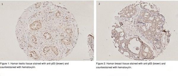 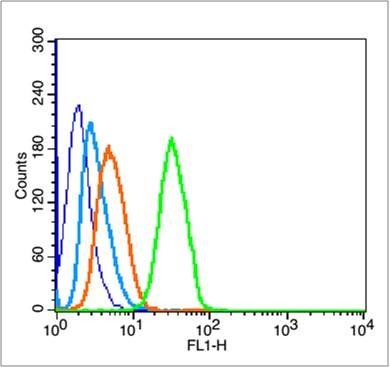 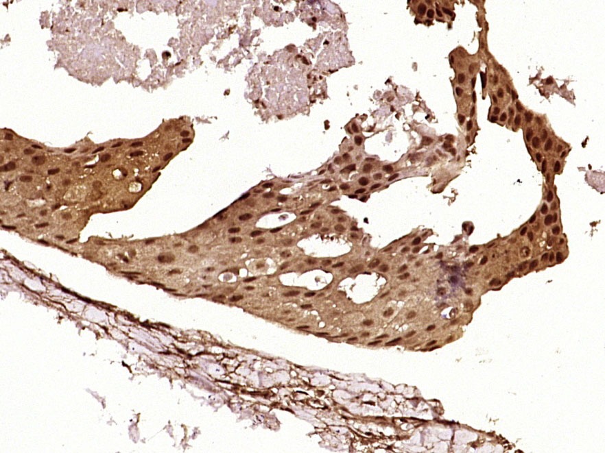 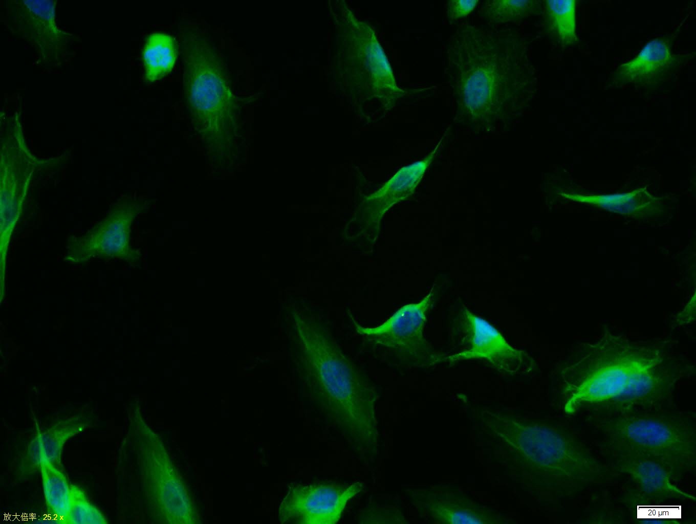 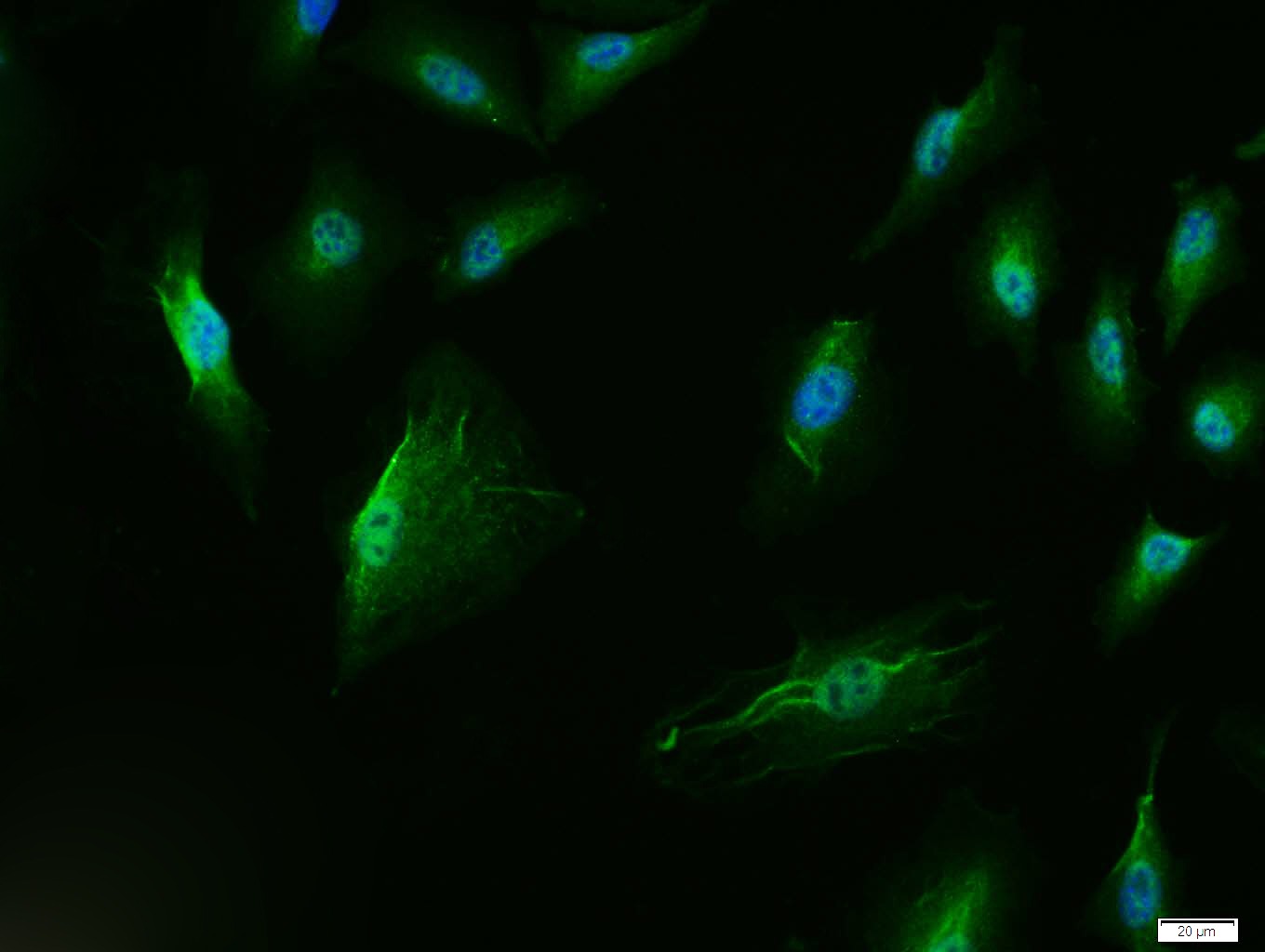 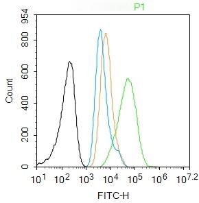 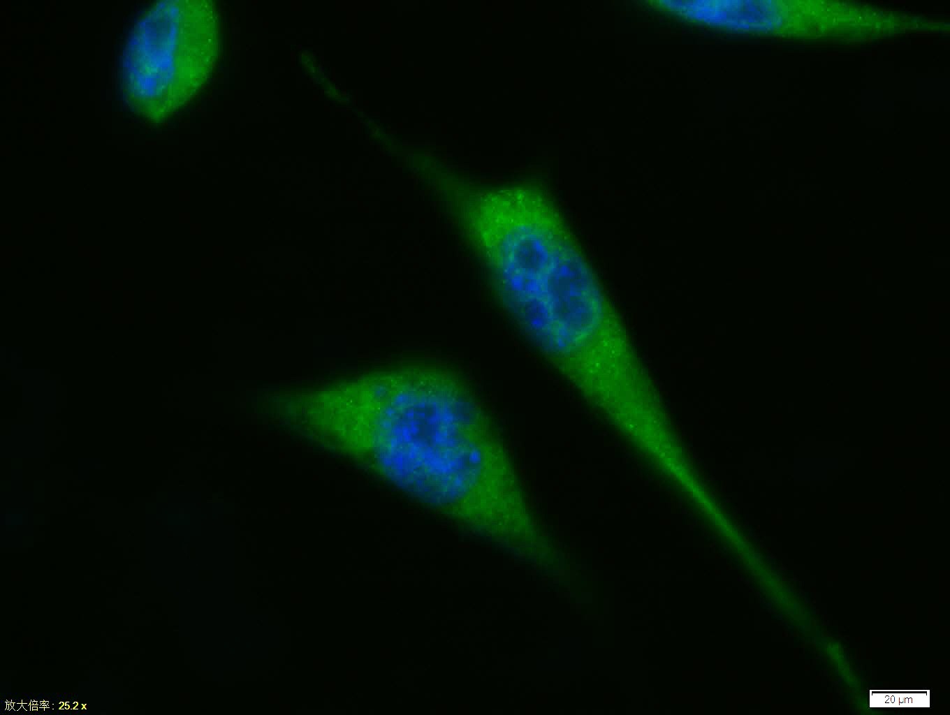 |
| Application | |
| Recommended Dose | WB: 1:500-2000; IHC-P: 1:100-500; IHC-Fr: 1:100-500; ICC/IF: 1:100; IF: 1:100-500; FCM: 1μg/Test |
| Antibody Type | Polyclonal |
| Host Species | Rabbit |
| Subcellular Localization | Cytoplasm. Nucleus. Nucleus, PML body. Endoplasmic reticulum. Note=Interaction with BANP promotes nuclear localization. Recruited into PML bodies together with CHEK2. Isoform 1: Nucleus. Cytoplasm. Note=Predominantly nuclear but localizes to the cytoplasm when expressed with isoform 4. Isoform 2: Nucleus. Cytoplasm. Note=Localized mainly in the nucleus with minor staining in the cytoplasm. Isoform 3: Nucleus. Cytoplasm. Note=Localized in the nucleus in most cells but found in the cytoplasm in some cells. Isoform 4: Nucleus. Cytoplasm. Note=Predominantly nuclear but translocates to the cytoplasm following cell stress. Isoform 7: Nucleus. Cytoplasm. Note=Localized mainly in the nucleus with minor staining in the cytoplasm. Isoform 8: Nucleus. Cytoplasm. Note=Localized in both nucleus and cytoplasm in most cells. In some cells, forms foci in the nucleus that are different from nucleoli. Isoform 9: Cytoplasm. |
| Tissue Specificity | Ubiquitous. Isoforms are expressed in a wide range of normal tissues but in a tissue-dependent manner. Isoform 2 is expressed in most normal tissues but is not detected in brain, lung, prostate, muscle, fetal brain, spinal cord and fetal liver. Isoform 3 |
| Construction | Polyclonal Antibody |
| Purification | Protein A purified |
| Appearance | Liquid |
| Formulation | 0.01M TBS (pH7.4) with 1% BSA, 0.02% Proclin300 and 50% Glycerol. |
| Concentration | 1 mg/mL |
| Research Background | This gene encodes a tumor suppressor protein containing transcriptional activation, DNA binding, and oligomerization domains. The encoded protein responds to diverse cellular stresses to regulate expression of target genes, thereby inducing cell cycle arrest, apoptosis, senescence, DNA repair, or changes in metabolism. Mutations in this gene are associated with a variety of human cancers, including hereditary cancers such as Li-Fraumeni syndrome. Alternative splicing of this gene and the use of alternate promoters result in multiple transcript variants and isoforms. Additional isoforms have also been shown to result from the use of alternate translation initiation codons (PMIDs: 12032546, 20937277). [provided by RefSeq, Feb 2013]. |
| Immunogen | KLH conjugated synthetic peptide: human P53 |
| Antigen Species | Human |
| Gene Name | TP53 |
| Gene ID | |
| Protein Name | Cellular tumor antigen p53 |
| Uniprot ID | |
| Biology Area | p53 pathway,p53 pathway,p53 Pathway,p53,p53,p53,Tumor Suppressors |
| Function | Acts as a tumor suppressor in many tumor types; induces growth arrest or apoptosis depending on the physiological circumstances and cell type. Involved in cell cycle regulation as a trans-activator that acts to negatively regulate cell division by controlling a set of genes required for this process. One of the activated genes is an inhibitor of cyclin-dependent kinases. Apoptosis induction seems to be mediated either by stimulation of BAX and FAS antigen expression, or by repression of Bcl-2 expression. Implicated in Notch signaling cross-over. Prevents CDK7 kinase activity when associated to CAK complex in response to DNA damage, thus stopping cell cycle progression. Isoform 2 enhances the transactivation activity of isoform 1 from some but not all TP53-inducible promoters. Isoform 4 suppresses transactivation activity and impairs growth suppression mediated by isoform 1. Isoform 7 inhibits isoform 1-mediated apoptosis. |
| Molecular Weight | Theoretical: 43 kDa. |
| Stability & Storage | Store at -20°C or -80°C for 12 months. Avoid repeated freeze-thaw cycles. |
| Transport | Shipping with blue ice. |

| Size | Quantity | Unit Price | Amount | Operation |
|---|
 Hello! How can I help you today?
 Copyright © 2015-2025 TargetMol Chemicals Inc. All Rights Reserved.

 For example, your dosage is 10 mg/kg Each animal weighs 20 g, and the dosage volume is 100 μL .