
Shopping Cart
Remove All Your shopping cart is currently empty

Synonyms: PERB11.1, MIC-A, MHC class I polypeptide-related sequence A, MGC21250, MGC111087, FLJ60820, FLJ36918, DAMA-345G11.2
Anti-MICA Polyclonal Antibody
| Pack Size | Price | USA Stock | Global Stock | Quantity |
|---|---|---|---|---|
| 50 µL | $222 | 7-10 days | 7-10 days | |
| 100 µL | $372 | 7-10 days | 7-10 days | |
| 200 µL | $528 | 7-10 days | 7-10 days |
| Description | Anti-MICA Polyclonal Antibody is a Rabbit antibody targeting MICA. Anti-MICA Polyclonal Antibody can be used in FCM,IF,IHC-Fr,IHC-P,WB. |
| Synonyms | PERB11.1, MIC-A, MHC class I polypeptide-related sequence A, MGC21250, MGC111087, FLJ60820, FLJ36918, DAMA-345G11.2 |
| Ig Type | IgG |
| Reactivity | Human,Mouse |
| Verified Activity | 1. Tissue/cell: human lung carcinoma; 4% Paraformaldehyde-fixed and paraffin-embedded; Antigen retrieval: citrate buffer (0.01M, pH6.0), Boiling bathing for 15 min; Block endogenous peroxidase by 3% Hydrogen peroxide for 30 min; Blocking buffer (normal goat serum) at 37°C for 20 min; Incubation: Anti-MICA/MHC I a Polyclonal Antibody, Unconjugated (TMAB-01136) 1:200, overnight at 4°C, followed by conjugation to the secondary antibody and DAb staining. 2. Tissue/cell: mouse intestine tissue; 4% Paraformaldehyde-fixed and paraffin-embedded; Antigen retrieval: citrate buffer (0.01M, pH6.0), Boiling bathing for 15 min; Block endogenous peroxidase by 3% Hydrogen peroxide for 30 min; Blocking buffer (normal goat serum) at 37°C for 20 min; Incubation: Anti-MICA/MHC I a Polyclonal Antibody, Unconjugated (TMAB-01136) 1:200, overnight at 4°C, followed by conjugation to the secondary antibody and DAb staining. 3. Tissue/cell: mouse lung tissue; 4% Paraformaldehyde-fixed and paraffin-embedded; Antigen retrieval: citrate buffer (0.01M, pH6.0), Boiling bathing for 15 min; Block endogenous peroxidase by 3% Hydrogen peroxide for 30 min; Blocking buffer (normal goat serum) at 37°C for 20 min; Incubation: Anti-MICA/MHC I a Polyclonal Antibody, Unconjugated (TMAB-01136) 1:200, overnight at 4°C, followed by conjugation to the secondary antibody and DAb staining. 4. Blank control (blue line): A431 (blue). Primary Antibody (green line): Rabbit Anti-MICA antibody (TMAB-01136) Dilution: 1 μg/10^6 cells; Isotype Control Antibody (orange line): Rabbit IgG. Secondary Antibody (white blue line): Goat anti-rabbit IgG-FITC Dilution: 1 μg/test. Protocol The cells were fixed with 2% paraformaldehyde (10 min), then permeabilized with 90% ice-cold methanol for 30 min on ice. Cells stained with Primary Antibody for 30 min at room temperature. The cells were then incubated in 1 X PBS/2% BSA/10% goat serum to block non-specific protein-protein interactions followed by the antibody for 15 min at room temperature. The secondary antibody used for 40 min at room temperature. 5. Sample: Lane 1: Mouse Large intestine tissue lysates Lane 2: Mouse Lung tissue lysates Lane 3: Mouse Kidney tissue lysates Lane 4: Mouse Liver tissue lysates Primary: Anti-MICA (TMAB-01136) at 1/1000 dilution Secondary: IRDye800CW Goat Anti-Rabbit IgG at 1/20000 dilution Predicted band size: 43 kDa Observed band size: 60 kDa 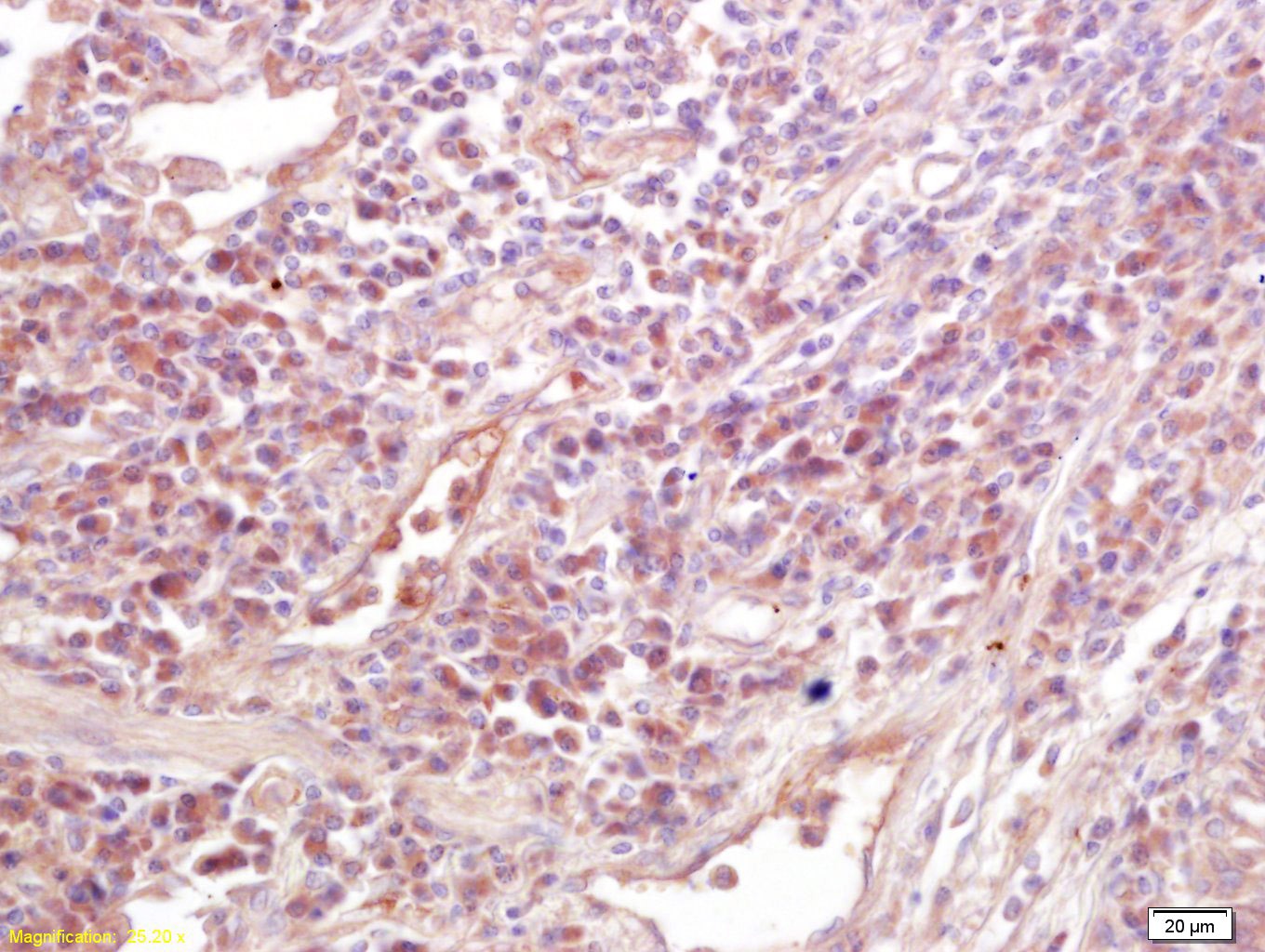 , ,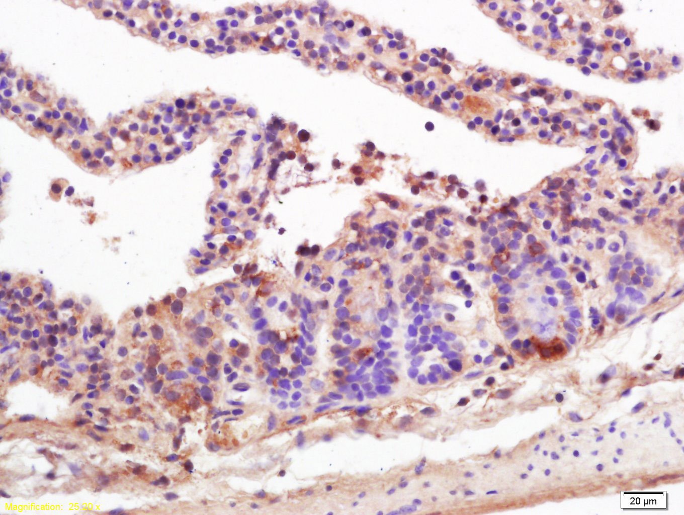 , ,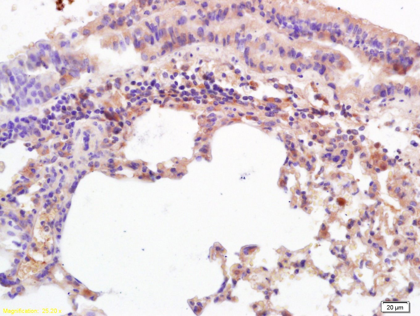 , ,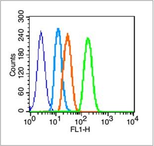 , ,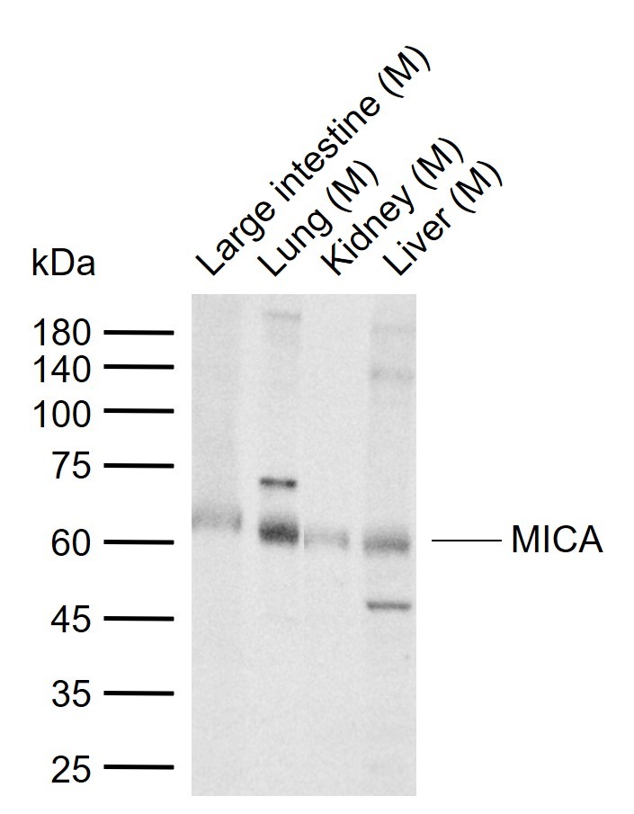 |
| Application | |
| Recommended Dose | WB: 1:500-2000; IHC-P: 1:100-500; IHC-Fr: 1:100-500; IF: 1:100-500; FCM: 1μg/Test |
| Antibody Type | Polyclonal |
| Host Species | Rabbit |
| Subcellular Localization | Cell membrane. Cytoplasm. Expressed on the cell surface in gastric epithelium, endothelial cells and fibroblasts and in the cytoplasm in keratinocytes and monocytes. Infection with human adenovirus 5 suppresses cell surface expression due to the adenoviral E3-19K protein which causes retention in the endoplasmic reticulum. |
| Tissue Specificity | Widely expressed with the exception of the central nervous system where it is absent. Expressed predominantly in gastric epithelium and also in monocytes, keratinocytes, endothelial cells, fibroblasts and in the outer layer of Hassal's corpuscles within t |
| Construction | Polyclonal Antibody |
| Purification | Protein A purified |
| Appearance | Liquid |
| Formulation | 0.01M TBS (pH7.4) with 1% BSA, 0.02% Proclin300 and 50% Glycerol. |
| Concentration | 1 mg/mL |
| Research Background | The MHC class I chain-related (MIC) proteins are related to the Major histocompatibility complex (MHC) class I proteins which are ubiquitously expressed and mediate the recognition of intracellular antigens by cytotoxic T cells. The MHC class I chain-related (MIC) proteins are recognized by NKG2D, a receptor on NK and T cells, and promote anti-tumor activity. MICA, a member of the MIC family, is widely expressed on many tumors, and it is the MICA/NKG2D interaction that is thought to stimulate the anti-tumor reactivity by T lymphocytes. MICA is present in virtually every tissue except the nervous system, suggesting that MIC protein expression may only be one component of the anti-tumor activity of the immune system. |
| Immunogen | KLH conjugated synthetic peptide: human MICA |
| Antigen Species | Human |
| Gene Name | MICA |
| Gene ID | |
| Protein Name | MHC class I chain-related protein A |
| Uniprot ID | |
| Biology Area | Class I,NK Cells |
| Function | Seems to have no role in antigen presentation. Acts as a stress-induced self-antigen that is recognized by gamma delta T-cells. Ligand for the KLRK1/NKG2D receptor. Binding to KLRK1 leads to cell lysis. |
| Molecular Weight | Theoretical: 43 kDa. Actual: 60 kDa. |
| Stability & Storage | Store at -20°C or -80°C for 12 months. Avoid repeated freeze-thaw cycles. |
| Transport | Shipping with blue ice. |
| Size | Quantity | Unit Price | Amount | Operation |
|---|
 Hello! How can I help you today?
 Copyright © 2015-2026 TargetMol Chemicals Inc. All Rights Reserved.
