
Shopping Cart
Remove All Your shopping cart is currently empty

Synonyms: low density lipoprotein receptor, LDLCQ2, LDL Receptor, LDL R, FHC, FH
Anti-LDLR Polyclonal Antibody
| Pack Size | Price | USA Stock | Global Stock | Quantity |
|---|---|---|---|---|
| 50 µL | $222 | 7-10 days | 7-10 days | |
| 100 µL | $374 | 7-10 days | 7-10 days | |
| 200 µL | $529 | 7-10 days | 7-10 days |
| Description | Anti-LDLR Polyclonal Antibody is a Rabbit antibody targeting LDLR. Anti-LDLR Polyclonal Antibody can be used in FCM, ICC/IF, WB. |
| Synonyms | low density lipoprotein receptor, LDLCQ2, LDL Receptor, LDL R, FHC, FH |
| Ig Type | IgG |
| Reactivity | Human,Mouse (predicted:Rat,Dog,Pig,Cow,Horse,Rabbit,GuineaPig) |
| Verified Activity | 1. Sample: Human colon Lysate at 45 μg; Primary: Anti-LDL receptor (TMAB-01057) at 1:300 dilution; Secondary: HRP conjugated Goat Anti-Rabbit Igg (secondary antibody) at 1: 5000 dilution; Predicted band size: 92 kDa Observed band size: 92 kDa 2. Sample: K562 (Human) Cell Lysate at 30 μg Raw264.7 (Mouse) Cell Lysate at 30 μg HepG2 (Human) Cell Lysate at 30 μg SW480 (Human) Cell Lysate at 30 μg Primary: Anti-LDL receptor (TMAB-01057) at 1/1000 dilution Secondary: IRDye800CW Goat Anti-Rabbit IgG at 1/20000 dilution Predicted band size: 92 kDa Observed band size: 120 kDa 3. Blank control: Raw264.7. Primary Antibody (green line): Rabbit Anti-LDL receptor antibody (TMAB-01057) Dilution: 1 μg/10^6 cells; Isotype Control Antibody (orange line): Rabbit IgG. Secondary Antibody: Goat anti-rabbit IgG-AF647 Dilution: 1 μg/test. Protocol The cells were fixed with 4% PFA (10 min at room temperature) and then permeabilized with 0.1% PBST for 20 min at room temperature. The cells were then incubated in 5% BSA to block non-specific protein-protein interactions for 30 min at room temperature. Cells stained with Primary Antibody for 30 min at room temperature. The secondary antibody used for 40 min at room temperature. 4. HepG2 cell; 4% Paraformaldehyde-fixed; Triton X-100 at room temperature for 20 min; Blocking buffer (normal goat serum) at 37°C for 20 min; Antibody incubation with (LDL receptor) polyclonal Antibody, Unconjugated (TMAB-01057) 1:100, 90 minutes at 37°C; followed by a conjugated Goat Anti-Rabbit IgG antibody at 37°C for 90 minutes, DAPI (blue) was used to stain the cell nucleus. 5. Blank control (black line): HepG2 (black) (The cells were fixed with 2% paraformaldehyde (10 min), then permeabilized with PBST for 30 min on room temperature) Primary Antibody (green line): Rabbit Anti-LDLreceptor antibody (TMAB-01057); Dilution: 1 μg/10^6 cells; Isotype Control Antibody (orange line): Rabbit IgG. Secondary Antibody (white blue line): Goat anti-rabbit IgG-FITC;Dilution: 1 μg/test. 6. Blank control: A431. Primary Antibody (green line): Rabbit Anti-LDL receptor antibody (TMAB-01057), Dilution: 1 μg/10^6 cells. Isotype Control Antibody (orange line): Rabbit IgG. Secondary Antibody: Goat anti-rabbit IgG-AF647, Dilution: 1 μg/test. Protocol A431 cells were fixed with 4% PFA (10 min at room temperature) and then permeabilized with 0.1% PBST for 20 min at room temperature. The cells were then incubated in 5% BSA to block non-specific protein-protein interactions for 30 min at room temperature. Cells stained with Primary Antibody for 30 min at room temperature. The secondary antibody used for 40 min at room temperature. 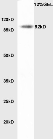 , ,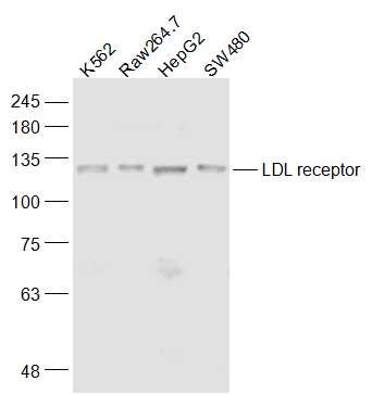 , ,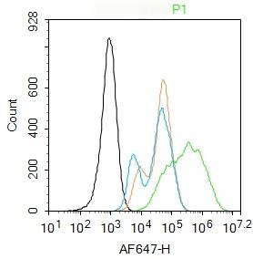 , ,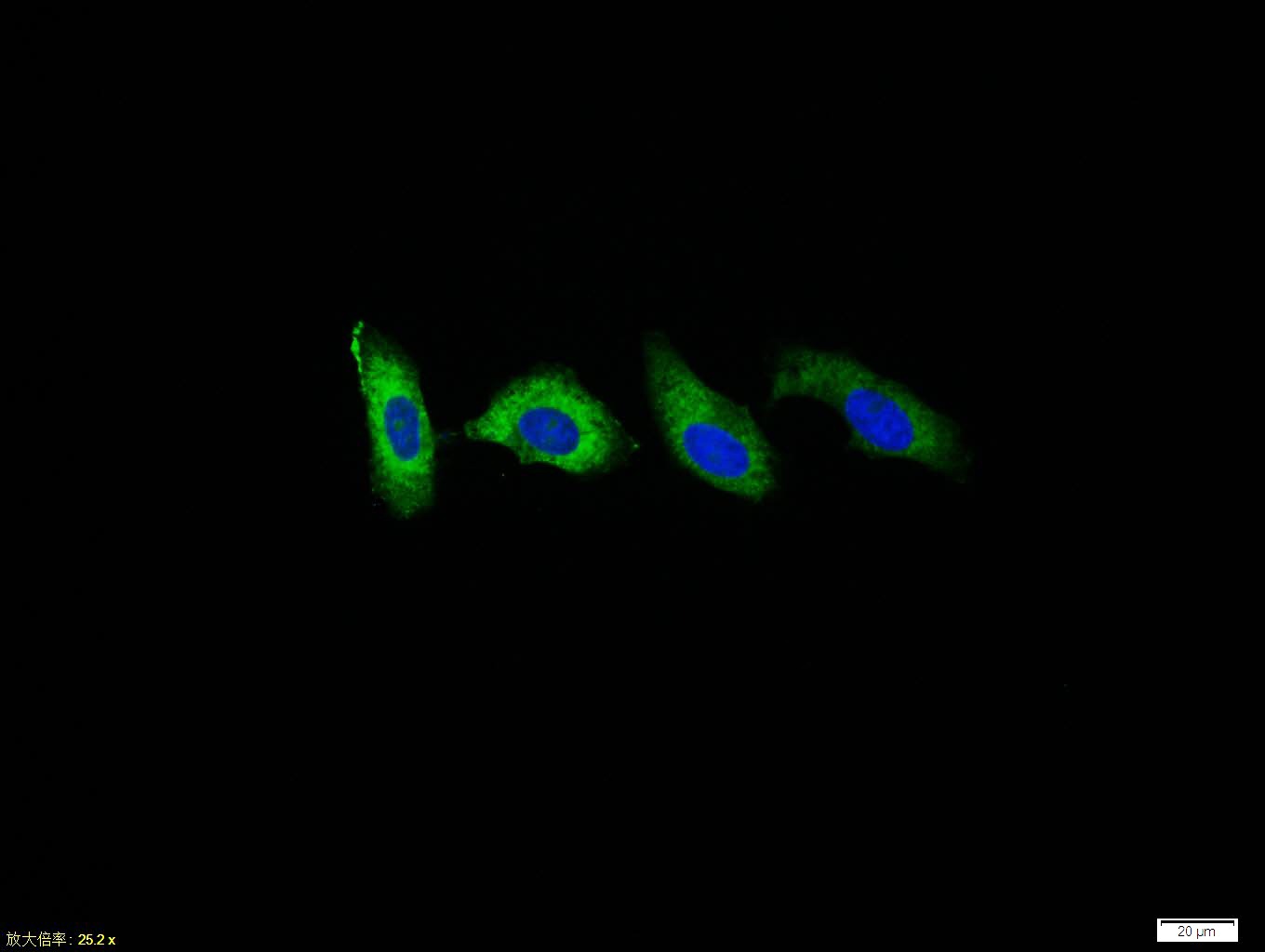 , ,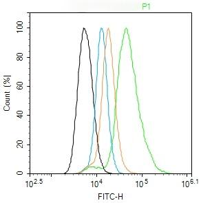 , ,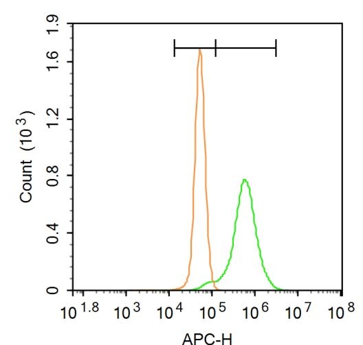 |
| Application | |
| Recommended Dose | FCM=1 μg/Test; ICC/IF=1:100-500; WB=1:500-2000 |
| Antibody Type | Polyclonal |
| Host Species | Rabbit |
| Subcellular Localization | Cell membrane; Single-pass type I membrane protein. Endomembrane system; Single-pass type I membrane protein. Membrane, clathrin-coated pit; Single-pass type I membrane protein. Note=Found distributed from the plasma membrane to intracellular compartments. |
| Tissue Specificity | Binds LDL, the major cholesterol-carrying lipoprotein of plasma, and transports it into cells by endocytosis. In order to be internalized, the receptor-ligand complexes must first cluster into clathrin-coated pits. In case of HIV-1 infection, functions as |
| Construction | Polyclonal Antibody |
| Purification | Protein A purified |
| Appearance | Liquid |
| Formulation | 0.01M TBS (pH7.4) with 1% BSA, 0.02% Proclin300 and 50% Glycerol. |
| Concentration | 1 mg/mL |
| Research Background | The low density lipoprotein receptor (LDLR) gene family consists of cell surface proteins involved in receptor-mediated endocytosis of specific ligands. The encoded protein is normally bound at the cell membrane, where it binds low density lipoprotein/cholesterol and is taken into the cell. Lysosomes release the cholesterol, which is made available for repression of microsomal enzyme 3-hydroxy-3-methylglutaryl coenzyme A (HMG CoA) reductase, the rate-limiting step in cholesterol synthesis. At the same time, a reciprocal stimulation of cholesterol ester synthesis takes place. Mutations in this gene cause the autosomal dominant disorder, familial hypercholesterolemia. Alternate splicing results in multiple transcript variants.[provided by RefSeq, May 2022] |
| Immunogen | KLH conjugated synthetic peptide: human LDL-R |
| Antigen Species | Human |
| Gene Name | LDLR |
| Gene ID | |
| Protein Name | Low-density lipoprotein receptor |
| Uniprot ID | |
| Biology Area | Metabolism of lipids and lipoproteins,Lipoprotein metabolism,Cholesterol Metabolism,Cholesterol Metabolism,Lipid metabolism,Lipoprotein metabolism,Cancer,Heart disease,Lipid metabolism,Lipid Messengers |
| Function | Binds LDL, the major cholesterol-carrying lipoprotein of plasma, and transports it into cells by endocytosis. In order to be internalized, the receptor-ligand complexes must first cluster into clathrin-coated pits. In case of HIV-1 infection, functions as a receptor for extracellular Tat in neurons, mediating its internalization in uninfected cells. |
| Molecular Weight | Theoretical: 92 kDa. Actual: 120 kDa. |
| Stability & Storage | Store at -20°C or -80°C for 12 months. Avoid repeated freeze-thaw cycles. |
| Transport | Shipping with blue ice. |
| Size | Quantity | Unit Price | Amount | Operation |
|---|
 Hello! How can I help you today?
 Copyright © 2015-2026 TargetMol Chemicals Inc. All Rights Reserved.
