
Shopping Cart
Remove All Your shopping cart is currently empty

Synonyms: XPE-binding factor, XPE-BF, XPE binding factor, XPE BF, XPE, XPCe, Xeroderma pigmentosum group E-complementing protein, Xeroderma pigmentosum group E complementing protein, XAP-1, XAP1, XAP 1, X associated protein 1, UV-DDB 1, UV-damaged DNA-binding protein 1, UV-damaged DNA-binding factor, UV DDB1, UV DDB 1, UV damaged DNA binding protein 1, UV damaged DNA binding factor, HBV X-associated protein 1, DNA damage-binding protein a, DNA damage-binding protein 1, DNA damage binding protein 1, DDBa, DDB1_HUMAN, Ddb1, DDB p127 subunit, DDB 1, Damage-specific DNA-binding protein 1, Damage specific DNA binding protein 1
Anti-DDB1 Antibody
(1Y859)
| Pack Size | Price | USA Stock | Global Stock | Quantity |
|---|---|---|---|---|
| 50 µL | $297 | 7-10 days | 7-10 days | |
| 100 µL | $497 | 7-10 days | 7-10 days |
| Description | Anti-DDB1 Antibody (1Y859) is a Rabbit antibody targeting DDB1. Anti-DDB1 Antibody (1Y859) can be used in ICC/IF,IHC,IP,WB. |
| Synonyms | XPE-binding factor, XPE-BF, XPE binding factor, XPE BF, XPE, XPCe, Xeroderma pigmentosum group E-complementing protein, Xeroderma pigmentosum group E complementing protein, XAP-1, XAP1, XAP 1, X associated protein 1, UV-DDB 1, UV-damaged DNA-binding protein 1, UV-damaged DNA-binding factor, UV DDB1, UV DDB 1, UV damaged DNA binding protein 1, UV damaged DNA binding factor, HBV X-associated protein 1, DNA damage-binding protein a, DNA damage-binding protein 1, DNA damage binding protein 1, DDBa, DDB1_HUMAN, Ddb1, DDB p127 subunit, DDB 1, Damage-specific DNA-binding protein 1, Damage specific DNA binding protein 1 |
| Ig Type | IgG |
| Clone | 1Y859 |
| Reactivity | Human,Mouse,Rat |
| Verified Activity | 1. Western blot analysis of DDB1 on different lysates using anti-DDB1 antibody at 1/500 dilution. Positive control: Lane 1: HepG2, Lane 2: NIH-3T3, Lane 3: MCF-7, Lane 4: Rat kidney tissue. 2. Immunohistochemical analysis of paraffin-embedded rat esophagus tissue using anti-DDB1 antibody. Counter stained with hematoxylin. 3. Immunohistochemical analysis of paraffin-embedded human liver cancer tissue using anti-DDB1 antibody. Counter stained with hematoxylin. 4. Immunohistochemical analysis of paraffin-embedded human kidney tissue using anti-DDB1 antibody. Counter stained with hematoxylin. 5. Immunohistochemical analysis of paraffin-embedded mouse brain tissue using anti-DDB1 antibody. Counter stained with hematoxylin. 6. ICC staining DDB1 in Hela cells (green). The nuclear counter stain is DAPI (blue). Cells were fixed in paraformaldehyde, permeabilised with 0.25% Triton X100/PBS. 7. ICC staining DDB1 in HUVEC cells (green). The nuclear counter stain is DAPI (blue). Cells were fixed in paraformaldehyde, permeabilised with 0.25% Triton X100/PBS. 8. ICC staining DDB1 in SH-SY5Y cells (green). The nuclear counter stain is DAPI (blue). Cells were fixed in paraformaldehyde, permeabilised with 0.25% Triton X100/PBS. 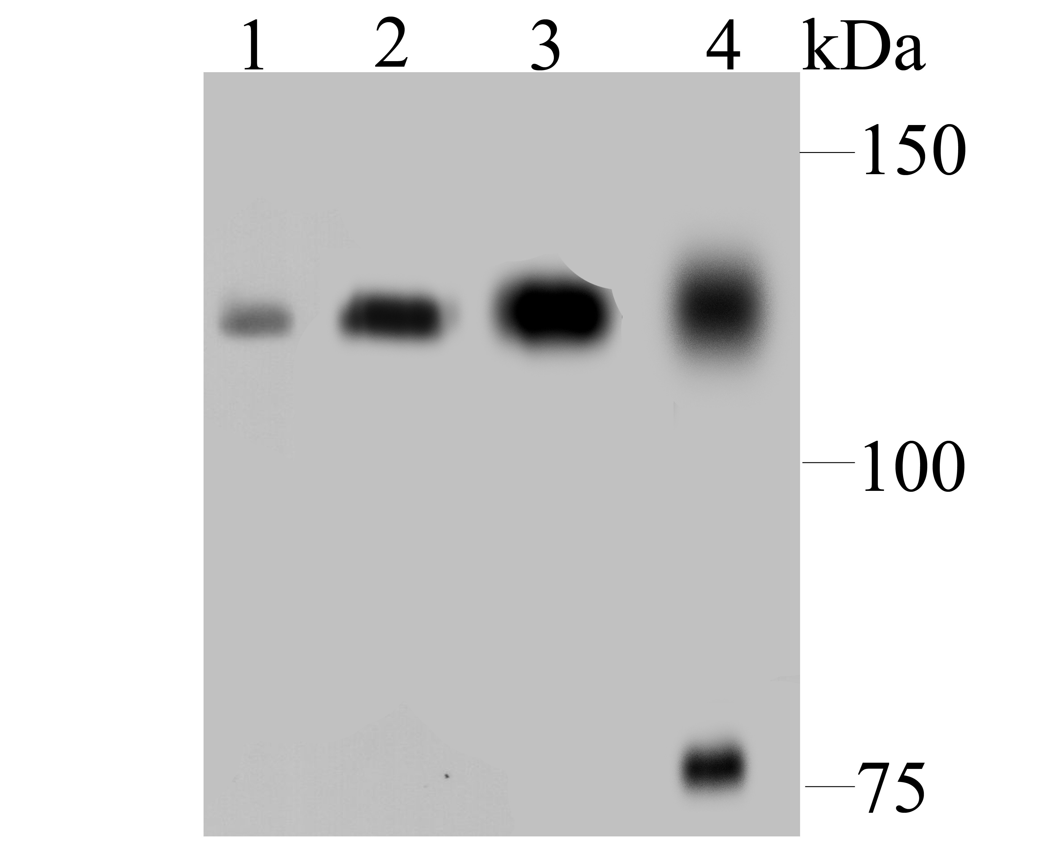 , ,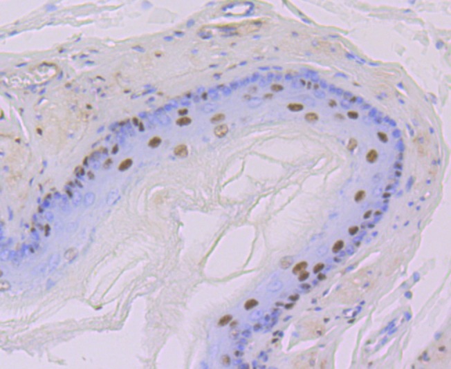 , ,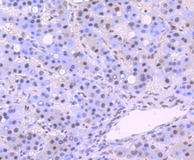 , ,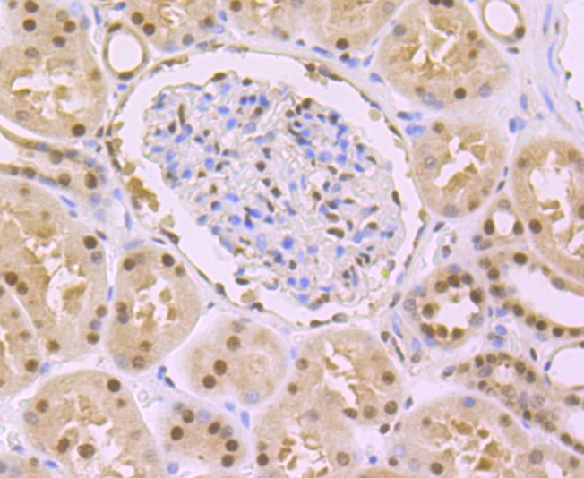 , ,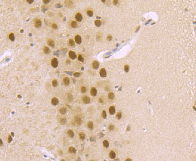 , ,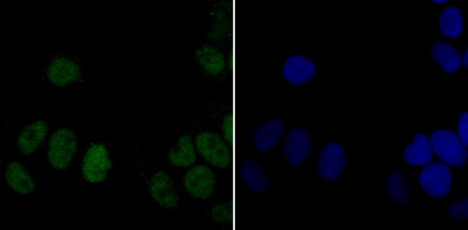 , ,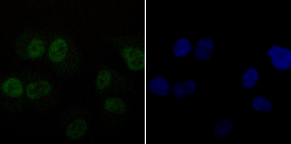 , ,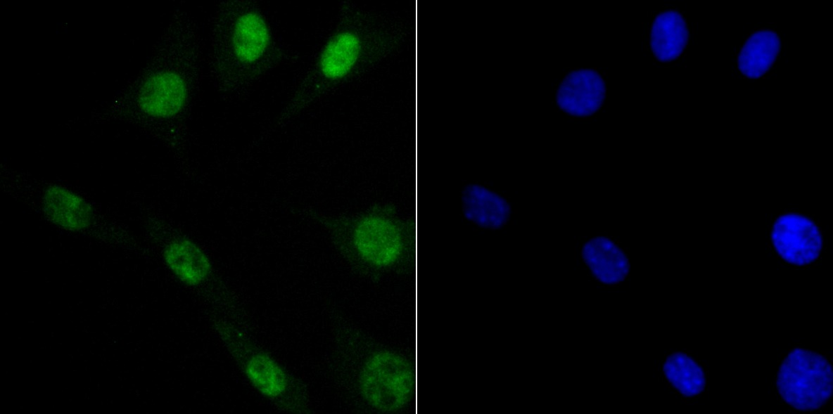 |
| Application | |
| Recommended Dose | WB: 1:500-2000; IHC: 1:50-200; ICC/IF: 1:50-200; IP: 1:10-50 |
| Antibody Type | Monoclonal |
| Host Species | Rabbit |
| Construction | Recombinant Antibody |
| Purification | ProA affinity purified |
| Appearance | Liquid |
| Formulation | 1*TBS (pH7.4), 1%BSA, 40%Glycerol. Preservative: 0.05% Sodium Azide. |
| Research Background | Damaged DNA binding protein (DDB) is a heterodimer composed of two subunits, p127 and p48, which are designated DDB1 and DDB2, respectively. The DDB heterodimer is involved in repairing DNA damaged by ultraviolet light. Specifically, DDB, also designated UV-damaged DNA binding protein (UV-DDB), xeroderma pigmentosum group E binding factor (XPE-BF) and hepatitis B virus X-associated protein 1 (XAP-1), binds to damaged cyclobutane pyrimidine dimers (CPDs). Mutations in the DDB2 gene are implicated as causes of xeroderma pigmentosum group E, an autosomal recessive disease in which patients are defective in nucleotide excision DNA repair. XPE is characterized by hypersensitivity of the skin to sunlight with a high frequency of skin cancer as well as neurologic abnormalities. The hepatitis B virus (HBV) X protein interacts with DDB1, which may mediate HBx transactivation. |
| Conjucates | Unconjugated |
| Immunogen | Recombinant Protein |
| Uniprot ID |
| Molecular Weight | Theoretical: 127 kDa. |
| Stability & Storage | Store at -20°C or -80°C for 12 months. Avoid repeated freeze-thaw cycles. |
| Transport | Shipping with blue ice. |
| Size | Quantity | Unit Price | Amount | Operation |
|---|
 Hello! How can I help you today?
 Copyright © 2015-2026 TargetMol Chemicals Inc. All Rights Reserved.
