
Shopping Cart
Remove All Your shopping cart is currently empty


Synonyms:
| Pack Size | Price | USA Stock | Global Stock | Quantity |
|---|---|---|---|---|
| 100 T | $155 | - | In Stock |
| Catalog No. | Product Name | Concentration | Solution | Packing |
|---|---|---|---|---|
| C0174-1 | JC-1 | 400 μM | DMSO | 100 μL × 5 |
| C0174-2 | CCCP | 10 mM | DMSO | 20 μL |
1.High sensitivity.
2.Well-established control system.
3.Low cytotoxicity.
4.Excellent probe stability.
5.Good compatibility, allowing co-staining with multiple fluorescent probes (e.g., Annexin V-FITC).
1.Detection of mitochondrial membrane potential changes during early apoptosis
2.Development of mitochondria-targeted therapeutics
3.Research on mitochondrial function and metabolic diseases
4.Studies on mitochondrial functional decline in aging cells
Dilute the stock solution with an appropriate diluent (serum-free culture medium or PBS) to prepare a working solution at a concentration of 2 μM.
If this kit is used with a 6-well plate, using a working solution at 20 μM and a detection volume of 1 mL per well, it allows for 100 assays. If used with a 96-well plate, using a working solution at 20 μM and a detection volume of 100 μL per well, it allows for up to 1000 assays.
1. Control Group Setup
(1) Positive Control Group (CCCP group):
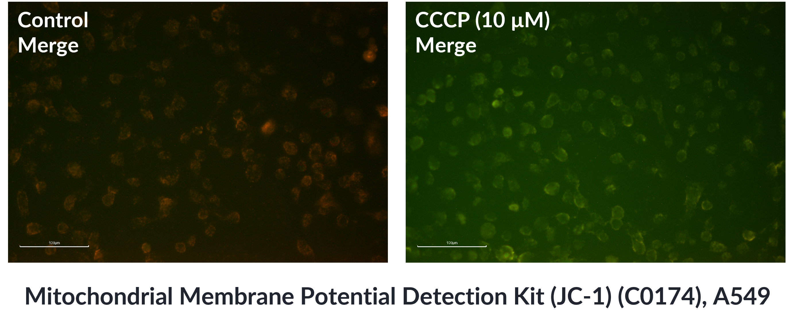
CCCP is a commonly used uncoupling agent. As a proton carrier, it transports protons across the inner mitochondrial membrane, thereby dissipating the proton gradient on both sides of the membrane. Since mitochondrial membrane potential depends on the maintenance of this proton gradient, CCCP treatment leads to a rapid decrease in mitochondrial membrane potential. Therefore, CCCP is widely used as a positive control in mitochondrial membrane potential assays.
Procedure:
Dilute the CCCP stock solution (10 mM) with serum-free culture medium to prepare a 10 µM CCCP working solution. Remove the old culture medium and add the CCCP working solution to the cells, then incubate for 20–30 min. After treatment, proceed with JC-1 staining of suspension or adherent cells as described below by adding an appropriate amount of JC-1 working solution for mitochondrial membrane potential detection.
Treatment with 10 µM CCCP for 20–30 min can induce loss of mitochondrial membrane potential in most cells. After JC-1 staining, cells with depolarized mitochondria exhibit green fluorescence, whereas cells with normal mitochondrial membrane potential show red fluorescence.
(2) Negative Control Group (Vehicle Control):
No drug treatment or other intervention is applied. This control is used to exclude non-specific fluorescence interference caused by the drug or experimental procedures.
(3) Blank Control Group:
Cells are neither treated with drugs nor stained with JC-1. This group is used to assess autofluorescence or background signals.
2. For Suspension Cells
(1) Centrifuge the cell suspension at 600 × g for 4 min at 4°C and discard the supernatant. Resuspend the cells in PBS, centrifuge again at 600 × g for 4 min at 4°C, discard the supernatant, and collect the cell pellet.
(2) Add an appropriate volume of JC-1 staining working solution to resuspend the cells and adjust the cell density to 5 × 10⁵–1 × 10⁶ cells/mL. Incubate the cells at 37°C for 20 min in the dark.
(3) After incubation, centrifuge at 600 × g for 4 min at 4°C and discard the supernatant.
(4) Resuspend the cells in PBS, centrifuge at 600 × g for 4 min at 4°C, and discard the supernatant. Repeat this wash step once.
(5) Resuspend the cells in an appropriate volume of PBS and observe staining under a fluorescence microscope or confocal laser scanning microscope. Alternatively, analyze the results using a flow cytometer or fluorescence spectrophotometer.
3. For Adherent Cells
(1) Remove the culture medium and wash the cells once with PBS.
(2) Add an appropriate amount of JC-1 staining working solution and incubate the cells at 37c for 20 min in the dark.
(3) After incubation, remove the staining solution and wash the cells twice with PBS.
(4) Add an appropriate amount of PBS to cover the cells and observe under a fluorescence microscope or confocal laser scanning microscope.
Note:
If analysis by flow cytometry or fluorescence spectrophotometry is required for adherent cells, the cells can first be digested with trypsin. After collection and resuspension, proceed with mitochondrial membrane potential detection according to the protocol for suspension cells described above.
4. Result Detection
During fluorescence measurement, it is not necessary to set the excitation and emission wavelengths exactly at their maximum values.
For green fluorescence, the excitation wavelength can be set at 490 nm and the emission wavelength at 530 nm.
For red fluorescence, the excitation wavelength can be set at 525 nm and the emission wavelength at 590 nm.
Store at 20°C, protected from light, for 1 year.
1.Staining efficiency may vary depending on sample type and experimental conditions. It is recommended to optimize the JC-1 working solution concentration and staining time through preliminary experiments.
2.The optimal working concentration and treatment time of CCCP may also vary with different samples. These conditions should be optimized by pre-experiments. A recommended working concentration range is 1–20 µM.
3.The JC-1 fluorescent probe is suitable for detecting mitochondrial membrane potential in live cells, but not for use in fixed cells or fixed tissues.
4.JC-1 is a fluorescent dye and is prone to photobleaching upon light exposure. Therefore, all procedures should be performed in the dark whenever possible.
5.The product is for R&D use only, not for diagnostic procedures, food, drug, household or other uses.
6.Please wear a lab coat and disposable gloves.
| Size | Quantity | Unit Price | Amount | Operation |
|---|
 Hello! How can I help you today?
 Copyright © 2015-2026 TargetMol Chemicals Inc. All Rights Reserved.
