 Your shopping cart is currently empty


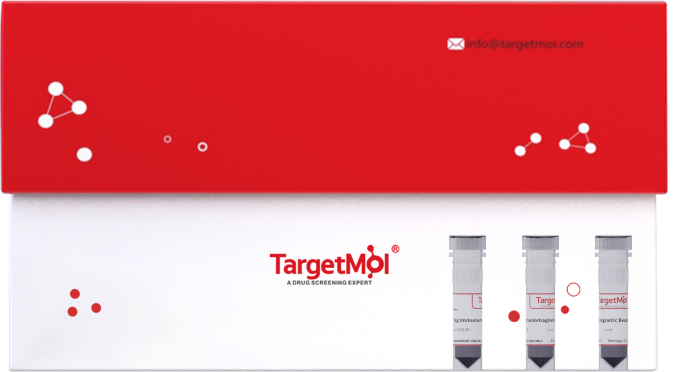
HA Tag Nanobody Immunomagnetic Beads
Nanobodies are variable domain fragments (VHH) derived from the naturally occurring heavy-chain antibodies of camelids (such as camels and alpacas). They are the smallest known natural functional antibody unit, typically about 12–15 kDa—around one-tenth the size of conventional IgG antibodies. Compared to traditional IgG antibodies, nanobodies are smaller in size, have higher affinity, and are free from light and heavy chain interference. They can bind closer to the epitope of the target protein, reducing steric hindrance. Additionally, they are less likely to dissociate, require milder elution conditions, and preserve higher biological activity of the samples.
| Pack Size | Price | USA Warehouse | Global Warehouse | Quantity |
|---|---|---|---|---|
| 500 μL | $350 | 7-10 days | 7-10 days | |
| 1 mL | $590 | 7-10 days | 7-10 days | |
| 5 mL | $2,845 | 7-10 days | 7-10 days |
 Product Information
| HA Tag Immunomagnetic Beads | Features |
|---|---|
| Bead Size | 2 μm |
| Binding Capacity | ≥ 1.5 mg HA Tag Protein/mL Beads |
| Storage Solution | 20 mM PBS, 5% BSA |
| Application | IP, Co-IP, ChIP, RIP |
 Features
1.No Antibody Chain Interference: Whether using denaturing or non-denaturing elution, the immunoprecipitated products are free from antibody heavy or light chains, facilitating downstream Western blot analysis.
2.Ultra-High Affinity: Exhibits nanomolar-level binding affinity, suitable for low-expression or hard-to-transfect cell samples.
3.Strong Binding Capacity: Directional coupling technology allows 10 μL of magnetic beads to bind approximately 15 μg of target recombinant protein.
4.High Specificity: Validated in over 10 blank cell lines with minimal non-specific adsorption.
5.Flexible Tag Recognition: Specifically binds to HA tags at either the N- or C-terminus of the bait protein, offering broad compatibility.
6.Heat Resistance: Tested at 45℃ for thermal stability, ensuring reliable performance during transportation and room-temperature storage.
 Prepare Reagents
| Reagent | Formulation |
|---|---|
| Washing Buffer (1×) | TBST: 50 mM Tris-HCl,150 mM NaCl, 0.1%(v/v) Tween-20,pH7.4 |
| HA Peptide Elution Buffer | PBS,1 mg/mL HA peptide (TP1276),pH 7.4 |
| Acidity Elution Buffer | 0.1M Glycine,0.1% (v/v) Tween-20,pH2.5 |
| Neutralization Buffer | 1 M Tris-HCl, pH 9.0 |
 Instructioins
Preparation of Cell Lysates
Select an appropriate lysis buffer to lyse cell samples and obtain cell lysates. Place on ice or store at -20℃ for long-term use.
Pretreatment of Magnetic Beads
1)Vortex for 1 min to resuspend the immunomagnetic beads. Take 20-30 µL of suspension and place it in a 1.5 mL EP tube.
2)Add 500 μL of Washing Buffer to the EP tube and gently invert several times to resuspend the beads. Keep the EP tube in a magnetic separator and stand for 1 min for magnetic separation. Finally, remove the supernatant and then take off the EP tube. Repeat the washing steps twice.
Immunoprecipitation
(1)Add 500 μL of prepared cell lysates to the EP tube. Place it on a rotating mixer and rotate at 37℃ for 30 min. For weak binding, incubate at room temperature for 1 hour or overnight at 4℃.
(2)After incubation, perform magnetic separation, then remove or save the supernatant for further analysis.
(3)Add 500 μL of Washing Buffer to the EP tube. Perform magnetic separation. Finally, remove the supernatant and then take off the EP tube. Repeat the washing steps 3 times.
Elution of Target Proteins
(1)Denaturing Elution: Suitable for SDS-PAGE detection. Add 100 μL of SDS-PAGE Loading Buffer to the EP tube. Mix well and heat at 95℃ for 5 min. Perform magnetic separation or centrifugation (room temperature, 13000 g, 10 min) to collect the supernatant.
(2)Neutral Elution: Add 50 μL of HA Peptide Elution Buffer to the EP tube. Incubate on a rotating mixer at 37℃ for 5-10 min (longer incubation time when below 37℃). Then perform magnetic separation or centrifugation to collect the supernatant.
(3)Acidity Elution: Add 100 μL of Acidity Elution Buffer to the EP tube. Incubate on a rotating mixer at 37℃ for 5-10 min. Perform magnetic separation or centrifugation to collect the supernatant. To adjust the pH of acidic elution buffer to neutral, add 50 μL of Neutralization Buffer to 100 μL elution.
Note: Nanobody-conjugated magnetic beads do not have heavy or light chain contamination issues. Denaturing elution is recommended as the first choice due to its higher elution efficiency.
 Storage
Store at 4℃ for 12 months.
 Precautions
1.Avoid freezing the beads. Store in solution to prevent drying.
2.The average magnetic separation time should be longer than 1 min.
3.Ensure uniform suspension by fully shaking the storage tube before use. Avoid bubbles during operation.
4.Use high-quality tips and test tubes to avoid sample loss due to adhesion.
5.Test the binding of proteins to beads by using the collected supernatant.
6.In IP experiments, the binding affinity of different proteins may vary. Users can select and prepare buffers according to experimental needs.
7.The product is for R&D use only, not for diagnostic procedures, food, drug, household or other uses.
8.Please wear a lab coat and disposable gloves.
 Instruction Manual
Keywords
| Size | Quantity | Unit Price | Amount | Operation |
|---|

Copyright © 2015-2026 TargetMol Chemicals Inc. All Rights Reserved.


