
Shopping Cart
Remove All Your shopping cart is currently empty

Synonyms:
| Pack Size | Price | USA Stock | Global Stock | Quantity |
|---|---|---|---|---|
| 100 T | $105 | - | In Stock |
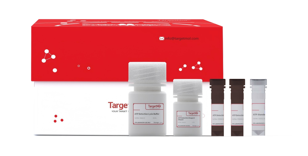
Taking 100T packing for example:
| Catalog No. | Product Name | Packing |
|---|---|---|
| C0212-1 | ATP Detection Reagent (10×) | 250 μL*2 |
| C0212-2 | ATP Detection Reagent Diluent | 5 mL |
| C0212-3 | ATP Standard Solution (0.5 mM) | 500 μL |
| C0212-4 | ATP Detection Lysis Buffer | 30 mL |
1.Simple Sample Preparation: Equipped with a dedicated lysis buffer that allows direct lysis of cells or tissue samples, eliminating the need for complex ATP extraction or boiling steps.
2.High Sensitivity Detection: Wide linear range with reliable detection from 0.1 nM to 10μM, meeting routine testing requirements for cell and tissue samples.
3.Stable Luminescent Signal: Minimal signal decay with stable and reliable readings. ATP standard curve results show that after 10 minutes of incubation, the chemiluminescent signal remains stable with no significant decrease for up to 120 minutes.
4.Good Sample Compatibility: Lysed samples can be used for both ATP measurement and subsequent protein-related experiments.
5.Efficient and Time-Saving: Fast detection workflow, enabling multi-sample analysis within 30~60 minutes.
Used for the quantitative detection of ATP levels in cell or tissue samples. It is widely applied in cell viability assessment, drug toxicity and efficacy screening, energy metabolism research, as well as studies related to cell injury and apoptosis.
**1. Sample Preparation (Recommended to perform at 4 ℃ or on ice)
1) Adherent Cells
After removing the culture medium, add an appropriate volume of lysis buffer according to the plate format (approximately 200 μL per well for a 6-well plate, 50 μL per well for a 96-well plate, and 25 μL per well for a 384-well plate).
Gently shake the plate or pipette up and down repeatedly to ensure the lysis buffer fully covers the cells and promotes complete lysis. Cells usually lyse rapidly upon contact with the lysis buffer.
After lysis, centrifuge at 12,000 × g for 5 minutes at 4°C. Collect the supernatant for subsequent ATP content measurement.
2) Suspension Cells
Centrifuge the cell suspension to collect the cell pellet. Discard the supernatant and gently resuspend the cells.
Add lysis buffer according to the same volume ratio as for adherent cells (approximately 200 μL per well for a 6-well plate, 50 μL per well for a 96-well plate, and 25 μL per well for a 384-well plate). Gently pipette up and down, flick the bottom of the tube, or briefly vortex to ensure complete lysis.
After lysis, centrifuge at 12,000 × g for 5 minutes at 4°C. Collect the supernatant for analysis.
3) Tissue Samples
Add lysis buffer at a ratio of approximately 100–200 μL per 20 mg of tissue. Homogenize thoroughly using a glass homogenizer or other appropriate homogenization equipment to ensure complete tissue lysis. After lysis, centrifuge at 12,000 × g for 5 minutes at 4°C, and collect the supernatant for subsequent analysis.
2. Preparation of Standard Solutions (Optional)
Thaw the ATP standard solution and related reagents slowly on ice. Dilute the ATP standard solution stepwise with ATP assay lysis buffer to prepare a series of ATP standards at different concentrations. The concentration range of the standards should be appropriately determined based on the expected ATP levels in the test samples. Generally, it is recommended to start at 10 μM and perform serial 3-fold dilutions to generate 8 concentration points for constructing a standard curve.
In subsequent experiments, the starting concentration and dilution range of the standards may be adjusted according to the actual ATP levels in the samples to obtain optimal linearity.
Note: When analyzing samples from 6-well plates, it is recommended to generate an ATP standard curve simultaneously to enable absolute quantification. For samples from 96-well or 384-well plates, if only relative ATP levels are being compared, preparation of a standard curve is optional depending on experimental requirements.
3. Preparation of ATP Detection Working Solution
Calculate the total volume of ATP detection working solution required according to the experimental design. It is recommended to use approximately 50 μL of ATP detection working solution per sample or standard.
Thaw the ATP detection reagent and related reagents slowly on ice. Take an appropriate amount of ATP detection reagent and dilute it with ATP detection reagent diluent at a ratio of 1:9 (v/v). Mix thoroughly to obtain the ATP detection working solution.
Note: The prepared ATP detection working solution is recommended to be used immediately. If short-term storage is required, keep it temporarily on ice.
4. ATP Assay
1) For samples from 6-well plates
a. Add 50 μL of ATP Detection Working Solution into each well of a white opaque 96-well plate or into designated assay tubes in advance. Incubate at room temperature for 5 minutes to deplete background ATP in the system and reduce baseline signal. For higher efficiency, the working solution may be added simultaneously to 10–20 wells or assay tubes at a time.
b. Add 50 μL of sample supernatant or ATP standard to each well or assay tube. Gently shake and mix for 5 minutes, then incubate at room temperature for 10 minutes protected from light.
After incubation, measure the signal using a luminometer or liquid scintillation counter. Record the RLU or CPM values. The recommended reading time is 1 second per well.
2) For samples in 96-well or 384-well plates
(Applicable when cells are directly cultured in white opaque plates)
a.Remove the culture plate from the incubator and allow it to equilibrate to room temperature.
Then add ATP lysis buffer to each well: 50 μL per well for a 96-well plate; 25 μL per well for a 384-well plate. Gently shake for 5 minutes to ensure complete cell lysis and ATP release.
b. Add the appropriate volume of ATP detection working solution to each well (50 μL per well for a 96-well plate; 25 μL per well for a 384-well plate). Shake gently for 5 minutes to mix thoroughly, then incubate at room temperature for 10 minutes in the dark. Measure the signal using a luminometer or liquid scintillation counter. The recommended detection time is 1 second per well.
Note:
Total volume per well in a 96-well plate: 100 μL culture medium + 50 μL ATP lysis buffer + 50 μL ATP detection working solution.
Total volume per well in a 384-well plate: 50 μL culture medium + 25 μL ATP lysis buffer + 25 μL ATP detection working solution.
3) Data Calculation
c. Calculate the ATP concentration in each sample according to the ATP standard curve.
d. To minimize the impact of differences in cell number or protein content among samples, it is recommended to determine the protein concentration using the BCA Protein Quantification Kit (C0050). The ATP content should then be normalized and expressed as nmol/mg protein for analysis.
Store at -20°C for up to 6 months; store at -80°C for up to 1 year.
C0212-1 should be protected from light.
1.The ATP detection reagent contains luciferase. Repeated freeze–thaw cycles may reduce enzyme activity. It is recommended to aliquot for further use. The diluted working solution should be freshly prepared before use and should not be stored frozen.
2.ATP is unstable after cell lysis. Sample preparation and detection are recommended to be performed at 4 °C or on ice. ATP can remain stable on ice for approximately 6 hours.
3.This kit requires a chemiluminescence instrument for detection. If unavailable, a liquid scintillation counter may also be used; however, the detection sensitivity depends on the performance of the instrument.
4.When using a multifunctional microplate reader capable of detecting chemiluminescence, opaque 96-well white or black plates are recommended to minimize well-to-well cross-talk and improve detection sensitivity.
5.If the detected ATP level in samples is significantly lower than expected, a portion of the sample may be boiled for 2 minutes after lysis and before centrifugation to ensure complete ATP release, followed by detection. Note that proteins in boiled samples are denatured and are not suitable for protein quantification, SDS-PAGE, or related experiments. For such analyses, use parallel samples that have not been boiled.
6.The product is for R&D use only, not for diagnostic procedures, food, drug, household or other uses.
7.Please wear a lab coat and disposable gloves.
| Size | Quantity | Unit Price | Amount | Operation |
|---|
 Hello! How can I help you today?
 Copyright © 2015-2026 TargetMol Chemicals Inc. All Rights Reserved.
