
Shopping Cart
- Remove All
 Your shopping cart is currently empty


Anti-TUBB3 Polyclonal Antibody is a Rabbit antibody targeting TUBB3. Anti-TUBB3 Polyclonal Antibody can be used in FCM,ICC/IF,IF,IHC-Fr,IHC-P,WB.
| Pack Size | Price | Availability | Quantity |
|---|---|---|---|
| 50 μL | $220 | 7-10 days | |
| 100 μL | $372 | 7-10 days | |
| 200 μL | $529 | 7-10 days |
| Description | Anti-TUBB3 Polyclonal Antibody is a Rabbit antibody targeting TUBB3. Anti-TUBB3 Polyclonal Antibody can be used in FCM,ICC/IF,IF,IHC-Fr,IHC-P,WB. |
| Synonyms | Tubulin beta-III, Tubulin beta-4 chain, Tubulin beta-3 chain, TUBB4, TUBB3 |
| Ig Type | IgG |
| Reactivity | Human,Mouse,Rat (predicted:Dog,Rabbit) |
| Verified Activity | 1. Tissue/cell: BV-2 cell; 4% Paraformaldehyde-fixed; Triton X-100 at room temperature for 20 min; Blocking buffer (normal goat serum) at 37°C for 20 min; Antibody incubation with (TUBB3) Polyclonal Antibody, Unconjugated (TMAB-01919) 1:200, 90 minutes at 37°C; followed by a conjugated Goat Anti-Rabbit IgG antibody at 37°C for 90 minutes, DAPI (5 μg/ml, blue) was used to stain the cell nucleus. 2. Blank control (blue): U-87MG Cells (fixed with 2% paraformaldehyde (10 min)). P rimary Antibody: Rabbit Anti-MGLUR3/AF647 Conjugated antibody (TMAB-01919/AF647), Dilution: 1 μg in 100 μL 1X PBS containing 0.5% BSA; Isotype Control Antibody: Rabbit IgG/AF647 (orange),used under the same conditions. 3. Paraformaldehyde-fixed, paraffin embedded (mouse cerebellum); Antigen retrieval by boiling in sodium citrate buffer (pH6.0) for 15 min; Block endogenous peroxidase by 3% hydrogen peroxide for 20 min; Blocking buffer (normal goat serum) at 37°C for 30 min; Antibody incubation with (TUBB3 (Neuronal Marker)) Polyclonal Antibody, Unconjugated (TMAB-01919) at 1:200 overnight at 4°C, followed by operating according to SP Kit (Rabbit) instructionsand DAB staining. 4. SH-SY5Y cell; 4% Paraformaldehyde-fixed; Ice-cold methanol at-20°C for 20 min; Blocking buffer (normal goat serum) at 37°C for 20 min; Antibody incubation with (TUBB3) polyclonal Antibody, Unconjugated (TMAB-01919) 1:100, 90 minutes at 37°C; followed by a FITC conjugated Goat Anti-Rabbit IgG antibody at 37°C for 90 minutes, DAPI (blue) was used to stain the cell nucleus. 5. Blank control: SH-SY5Y. Primary Antibody (green line): Rabbit Anti-TUBB3 (Neuronal Marker) antibody (TMAB-01919) Dilution: 1 μg/Test; Secondary Antibody: Goat anti-rabbit IgG-FITC Dilution: 0.5 μg/Test. Protocol The cells were fixed with 4% PFA (10 min at room temperature) and then permeabilized with 90% ice-cold methanol for 20 min at-20°C. The cells were then incubated in 5% BSA to block non-specific protein-protein interactions for 30 min at room temperature. Cells stained with Primary Antibody for 30 min at room temperature. The secondary antibody used for 40 min at room temperature. 6. Blank control: SH-SY5Y. Primary Antibody (green line): Rabbit Anti-TUBB3 (Neuronal Marker) antibody (TMAB-01919) Dilution: 1 μg/Test; Secondary Antibody: Goat anti-rabbit IgG-FITC Dilution: 0.5 μg/Test. Protocol The cells were fixed with 4% PFA (10 min at room temperature) and then permeabilized with 90% ice-cold methanol for 20 min at-20°C. The cells were then incubated in 5% BSA to block non-specific protein-protein interactions for 30 min at room temperature. Cells stained with Primary Antibody for 30 min at room temperature. The secondary antibody used for 40 min at room temperature. 7. Paraformaldehyde-fixed, paraffin embedded (Rat brain); Antigen retrieval by boiling in sodium citrate buffer (pH6.0) for 15 min; Block endogenous peroxidase by 3% hydrogen peroxide for 20 min; Blocking buffer (normal goat serum) at 37°C for 30 min; Antibody incubation with (TUBB3) Polyclonal Antibody, Unconjugated (TMAB-01919) at 1:400 overnight at 4°C, followed by a conjugated Goat Anti-Rabbit IgG antibody for 90 minutes, and DAPI for nucleus staining. 8. Paraformaldehyde-fixed, paraffin embedded (Mouse brain); Antigen retrieval by boiling in sodium citrate buffer (pH6.0) for 15 min; Block endogenous peroxidase by 3% hydrogen peroxide for 20 min; Blocking buffer (normal goat serum) at 37°C for 30 min; Antibody incubation with (TUBB3) Polyclonal Antibody, Unconjugated (TMAB-01919) at 1:400 overnight at 4°C, followed by a conjugated Goat Anti-Rabbit IgG antibody for 90 minutes, and DAPI for nucleus staining. 9. Sample: U251 (Human) Cell Lysate at 30 μg Primary: Anti-TUBB3 (Neuronal Marker) (TMAB-01919) at 1/1000 dilution Secondary: IRDye800CW Goat Anti-Rabbit IgG at 1/20000 dilution Predicted band size: 50-55 kDa Observed band size: 50 kDa 10. Sample: BV-2 (Rat) Cell Lysate at 30 μg Primary: Anti-TUBB3 (Neuronal Marker) (TMAB-01919) at 1/1000 dilution Secondary: IRDye800CW Goat Anti-Rabbit IgG at 1/20000 dilution Predicted band size: 50-55 kDa Observed band size: 50 kDa 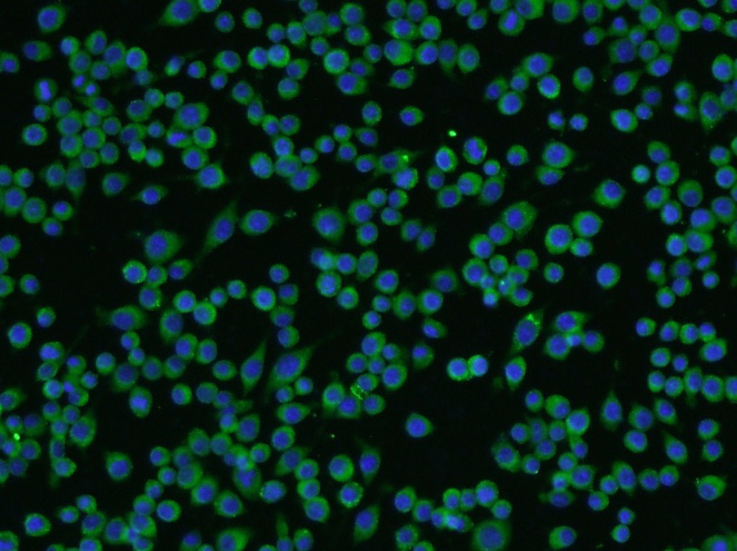 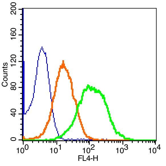 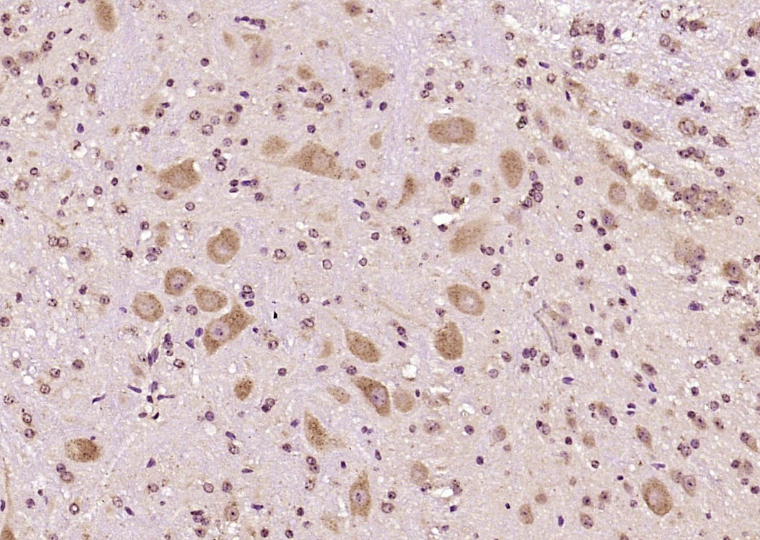 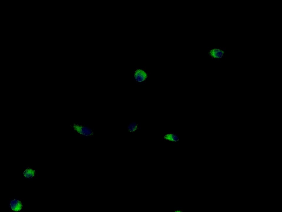 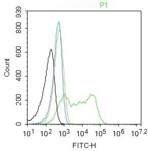 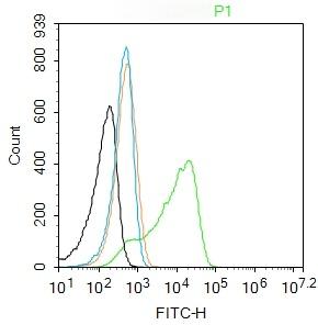 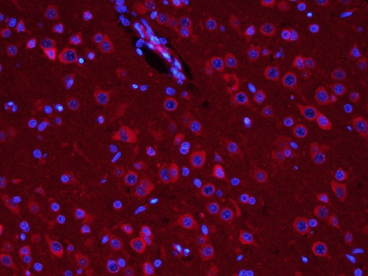 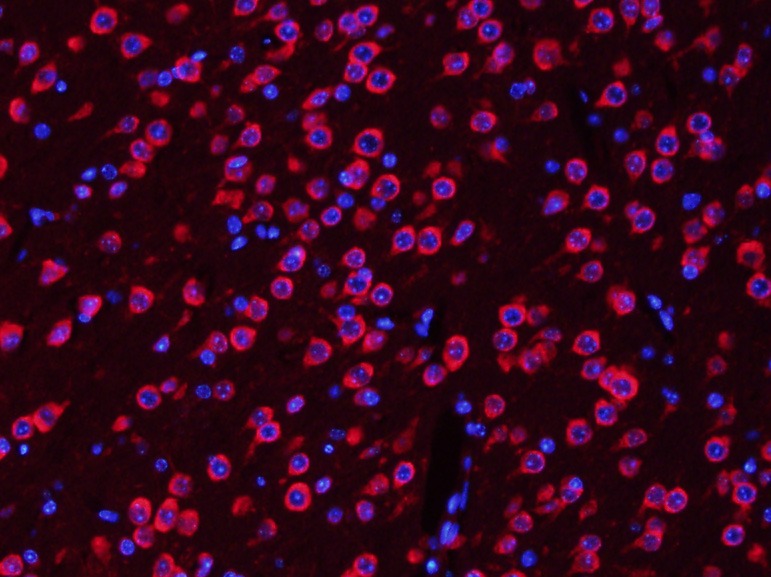 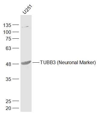 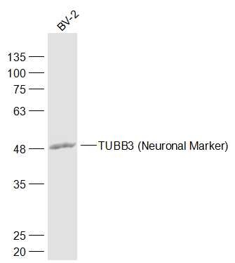 |
| Application | |
| Recommended Dose | WB: 1:500-2000; IHC-P: 1:100-500; IHC-Fr: 1:100-500; ICC/IF: 1:100; IF: 1:200-800; FCM: 1μg/Test |
| Antibody Type | Polyclonal |
| Host Species | Rabbit |
| Subcellular Localization | Cytoplasm, cytoskeleton. |
| Tissue Specificity | Expression is primarily restricted to centraland peripheral nervous system. Greatly increased expression in mostcancerous tissues. |
| Construction | Polyclonal Antibody |
| Purification | Protein A purified |
| Appearance | Liquid |
| Formulation | 0.01M TBS (pH7.4) with 1% BSA, 0.02% Proclin300 and 50% Glycerol. |
| Concentration | 1 mg/mL |
| Research Background | Neuronal Marker Beta III tubulin is abundant in the central and peripheral nervous systems (CNS and PNS) where it is prominently expressed during fetal and postnatal development. As exemplified in cerebellar and sympathoadrenal neurogenesis, the distribution of beta III is neuron-associated, exhibiting distinct temporospatial gradients according to the regional neuroepithelia of origin. However, transient expression of this protein is also present in the subventricular zones of the CNS comprising putative neuronal- and/or glial precursor cells, as well as in Kulchitsky neuroendocrine cells of the fetal respiratory epithelium. This temporally restricted, potentially non-neuronal expression may have implications in the identification of presumptive neurons derived from embryonic stem cells. |
| Immunogen | KLH conjugated synthetic peptide: human beta III Tubulin |
| Antigen Species | Human |
| Gene Name | TUBB3 |
| Gene ID | |
| Protein Name | Tubulin beta-3 chain |
| Uniprot ID | |
| Biology Area | Neuronal,Tubulin,Microtubules |
| Function | Tubulin is the major constituent of microtubules. Itbinds two moles of GTP, one at an exchangeable site on the betachain and one at a non-exchangeable site on the alpha chain. TUBB3plays a critical role in proper axon guidance and mantainance. |
| Molecular Weight | Theoretical: 50-55 kDa. |
| Stability & Storage | Store at -20°C or -80°C for 12 months. Avoid repeated freeze-thaw cycles. |
| Transport | Shipping with blue ice. |
 Hello! How can I help you today?
Copyright © 2015-2025 TargetMol Chemicals Inc. All Rights Reserved.
 For example, your dosage is 10 mg/kg Each animal weighs 20 g, and the dosage volume is 100 μL .