
Shopping Cart
Remove All Your shopping cart is currently empty

Synonyms: RIPK1, RIP1, RIP, Receptor-interacting serine/threonine-protein kinase 1, Receptor-interacting protein 1 (RIP-1), Cell death protein RIP
Anti-RIPK1 Polyclonal Antibody
| Pack Size | Price | USA Stock | Global Stock | Quantity |
|---|---|---|---|---|
| 50 µL | $220 | 7-10 days | 7-10 days | |
| 100 µL | $373 | 7-10 days | 7-10 days | |
| 200 µL | $529 | 7-10 days | 7-10 days |
| Description | Anti-RIPK1 Polyclonal Antibody is a Rabbit antibody targeting RIPK1. Anti-RIPK1 Polyclonal Antibody can be used in IF,IHC-Fr,IHC-P,WB. |
| Synonyms | RIPK1, RIP1, RIP, Receptor-interacting serine/threonine-protein kinase 1, Receptor-interacting protein 1 (RIP-1), Cell death protein RIP |
| Ig Type | IgG |
| Reactivity | Human,Mouse,Rat (predicted:Pig,Cow,Horse,Rabbit) |
| Verified Activity | 1. Paraformaldehyde-fixed, paraffin embedded (Rat brain); Antigen retrieval by microwave in sodium citrate buffer (pH6.0); Block endogenous peroxidase by 3% hydrogen peroxide for 30 minutes; Blocking buffer (3% BSA) at RT for 30 min; Antibody incubation with (RIPK-1) Polyclonal Antibody, Unconjugated (TMAB-01620) at 1:400 overnight at 4°C, followed by conjugation to the secondary antibody (labeled with HRP) and DAB staining. 2. Paraformaldehyde-fixed, paraffin embedded (Mouse brain); Antigen retrieval by microwave in sodium citrate buffer (pH6.0); Block endogenous peroxidase by 3% hydrogen peroxide for 30 minutes; Blocking buffer (3% BSA) at RT for 30 min; Antibody incubation with (RIPK-1) Polyclonal Antibody, Unconjugated (TMAB-01620) at 1:400 overnight at 4°C, followed by conjugation to the secondary antibody (labeled with HRP) and DAB staining. 3. Sample: Cerebrum (Mouse) Lysate at 40 μg Primary: Anti-RIPK-1 (TMAB-01620) at 1/1000 dilution Secondary: IRDye800CW Goat Anti-Rabbit IgG at 1/20000 dilution Predicted band size: 74 kDa Observed band size: 70 kDa 4. Sample: Raji (Human) Cell Lysate at 30 μg Primary: Anti-RIPK-1 (TMAB-01620) at 1/1000 dilution Secondary: IRDye800CW Goat Anti-Rabbit IgG at 1/20000 dilution Predicted band size: 74 kDa Observed band size: 70 kDa 5. Paraformaldehyde-fixed, paraffin embedded (human cervical carcinoma); Antigen retrieval by boiling in sodium citrate buffer (pH6.0) for 15 min; Block endogenous peroxidase by 3% hydrogen peroxide for 20 min; Blocking buffer (normal goat serum) at 37°C for 30 min; Incubation with (RIPK1) Polyclonal Antibody, Unconjugated (TMAB-01620) at 1:200 overnight at 4°C, followed by operating according to SP Kit (Rabbit) instructionsand DAB staining. 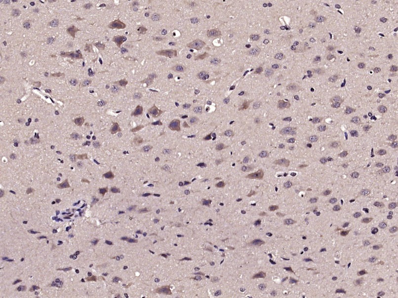 , ,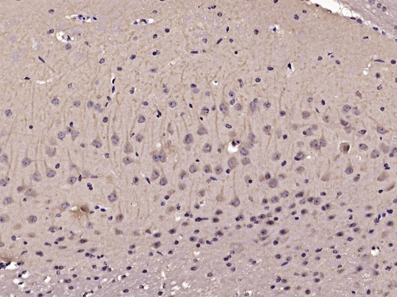 , ,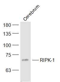 , , , ,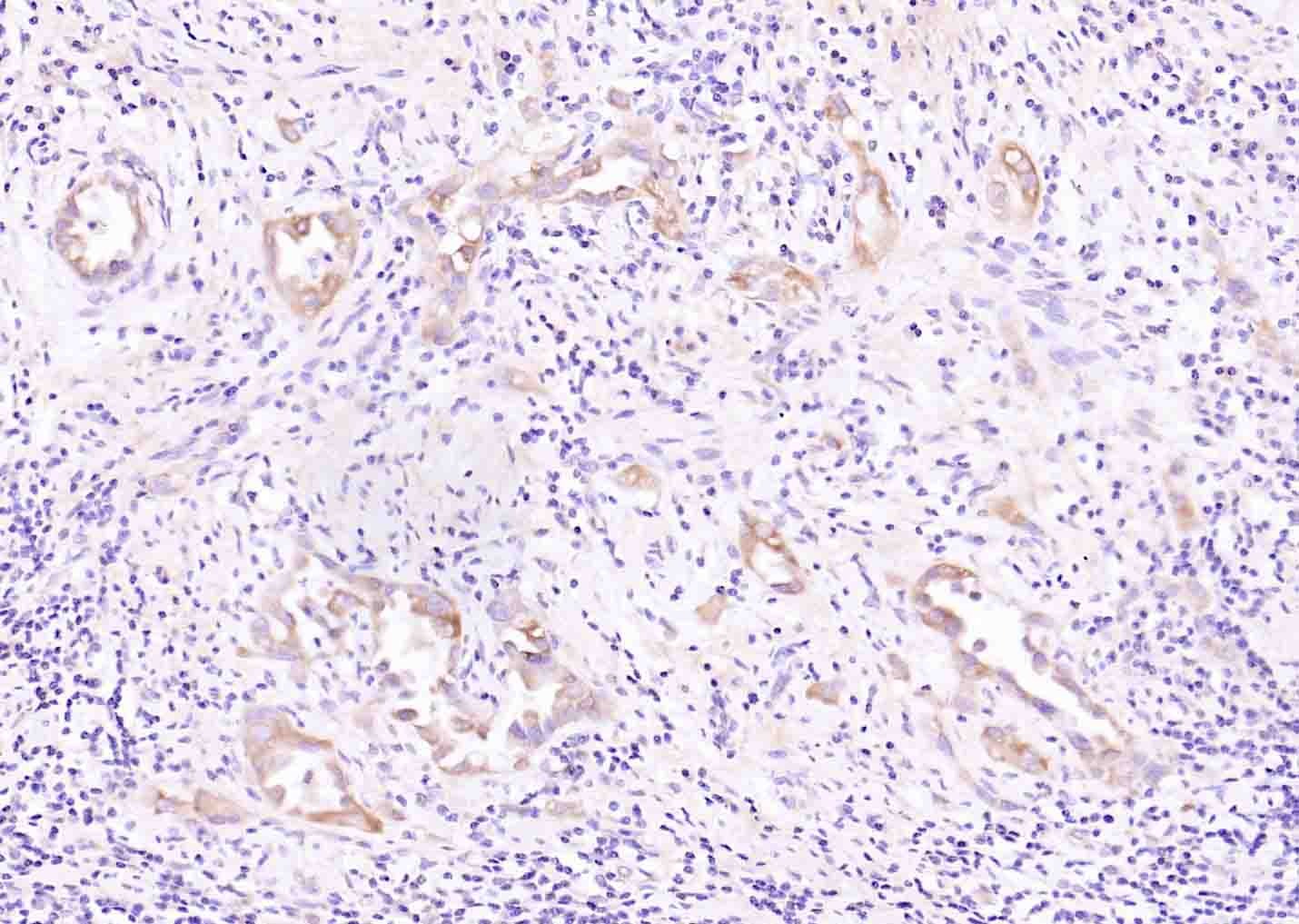 |
| Application | |
| Recommended Dose | WB: 1:500-2000; IHC-P: 1:100-500; IHC-Fr: 1:100-500; IF: 1:100-500 |
| Antibody Type | Polyclonal |
| Host Species | Rabbit |
| Subcellular Localization | Cytoplasm. Cell membrane. |
| Tissue Specificity | Expressed ubiquitously but at low levels. Shows a strong expression in the erythrocytes. |
| Construction | Polyclonal Antibody |
| Purification | Protein A purified |
| Appearance | Liquid |
| Formulation | 0.01M TBS (pH7.4) with 1% BSA, 0.02% Proclin300 and 50% Glycerol. |
| Concentration | 1 mg/mL |
| Research Background | Essential adapter molecule for the activation of NF-kappa-B. Following different upstream signals (binding of inflammatory cytokines, stimulation of pathogen recognition receptors, or DNA damage), particular RIPK1-containing complexes are formed, initiating a limited number of cellular responses. Upon TNFA stimulation RIPK1 is recruited to a TRADD-TRAF complex initiated by TNFR1 trimerization. There, it is ubiquitinated via 'Lys-63'-link chains, inducing its association with the IKK complex, and its activation through NEMO binding of polyubiquitin chains. |
| Immunogen | KLH conjugated synthetic peptide: human RIPK1 |
| Antigen Species | Human |
| Gene Name | RIPK1 |
| Gene ID | |
| Protein Name | Receptor-interacting serine/threonine-protein kinase 1 |
| Uniprot ID | |
| Function | Serine-threonine kinase which transduces inflammatory and cell-death signals (necroptosis) following death receptors ligation, activation of pathogen recognition receptors (PRRs), and DNA damage. Upon activation of TNFR1 by the TNF-alpha family cytokines, TRADD and TRAF2 are recruited to the receptor. Ubiquitination by TRAF2 via 'Lys-63'-link chains acts as a critical enhancer of communication with downstream signal transducers in the mitogen-activated protein kinase pathway and the NF-kappa-B pathway, which in turn mediate downstream events including the activation of genes encoding inflammatory molecules. Polyubiquitinated protein binds to IKBKG/NEMO, the regulatory subunit of the IKK complex, a critical event for NF-kappa-B activation. Interaction with other cellular RHIM-containing adapters initiates gene activation and cell death. RIPK1 and RIPK3 association, in particular, forms a necroptosis-inducing complex. |
| Molecular Weight | Theoretical: 74 kDa. Actual: 70 kDa. |
| Stability & Storage | Store at -20°C or -80°C for 12 months. Avoid repeated freeze-thaw cycles. |
| Transport | Shipping with blue ice. |
| Size | Quantity | Unit Price | Amount | Operation |
|---|
 Hello! How can I help you today?
 Copyright © 2015-2026 TargetMol Chemicals Inc. All Rights Reserved.
