
Shopping Cart
Remove All Your shopping cart is currently empty

Synonyms: Transforming growth factor-beta-activated kinase 1, TGF-beta-activated kinase 1, TGF1a, TGF beta activated kinase 1, TAK1 (p-Thr187), TAK1 (p-T187), TAK1, p-TAK1 (Thr187), p-TAK1 (T187), Mitogen-activated protein kinase kinase kinase 7, MEKK7, MAPKKK7, Map3k7, MAP3K 7, M3K7
Anti-Phospho-TAK1
(Thr187) Polyclonal Antibody
| Pack Size | Price | USA Stock | Global Stock | Quantity |
|---|---|---|---|---|
| 50 µL | $222 | 7-10 days | 7-10 days | |
| 100 µL | $372 | 7-10 days | 7-10 days | |
| 200 µL | $528 | 7-10 days | 7-10 days |
| Description | Anti-Phospho-TAK1 (Thr187) Polyclonal Antibody is a Rabbit antibody targeting Phospho-TAK1 (Thr187). Anti-Phospho-TAK1 (Thr187) Polyclonal Antibody can be used in FCM,IF,IHC-Fr,IHC-P,WB. |
| Synonyms | Transforming growth factor-beta-activated kinase 1, TGF-beta-activated kinase 1, TGF1a, TGF beta activated kinase 1, TAK1 (p-Thr187), TAK1 (p-T187), TAK1, p-TAK1 (Thr187), p-TAK1 (T187), Mitogen-activated protein kinase kinase kinase 7, MEKK7, MAPKKK7, Map3k7, MAP3K 7, M3K7 |
| Ig Type | IgG |
| Reactivity | Human,Mouse,Rat (predicted:Chicken,Pig,Cow,Horse,Rabbit) |
| Verified Activity | 1. Paraformaldehyde-fixed, paraffin embedded (Human stomach); Antigen retrieval by microwave in sodium citrate buffer (pH6.0); Block endogenous peroxidase by 3% hydrogen peroxide for 30 minutes; Blocking buffer (3% BSA) at RT for 30 min; Antibody incubation with (Phospho-TAK1 (Thr187)) Polyclonal Antibody, Unconjugated (TMAB-01515) at 1:400 overnight at 4°C, followed by conjugation to the secondary antibody (labeled with HRP) and DAB staining. 2. Paraformaldehyde-fixed, paraffin embedded (Mouse brain); Antigen retrieval by microwave in sodium citrate buffer (pH6.0); Block endogenous peroxidase by 3% hydrogen peroxide for 30 minutes; Blocking buffer (3% BSA) at RT for 30 min; Antibody incubation with (Phospho-TAK1 (Thr187)) Polyclonal Antibody, Unconjugated (TMAB-01515) at 1:400 overnight at 4°C, followed by conjugation to the secondary antibody (labeled with HRP) and DAB staining. 3. Sample: Muscle (Mouse) Lysate at 40 μg Primary: Anti-Phospho-TAK1 (Thr187) (TMAB-01515) at 1/1000 dilution Secondary: IRDye800CW Goat Anti-Rabbit IgG at 1/20000 dilution Predicted band size: 67 kDa Observed band size: 70 kDa 4. Paraformaldehyde-fixed, paraffin embedded (Rat brain); Antigen retrieval by microwave in sodium citrate buffer (pH6.0); Block endogenous peroxidase by 3% hydrogen peroxide for 30 minutes; Blocking buffer (3% BSA) at RT for 30 min; Antibody incubation with (Phospho-TAK1 (Thr187)) Polyclonal Antibody, Unconjugated (TMAB-01515) at 1:400 overnight at 4°C, followed by conjugation to the secondary antibody (labeled with HRP) and DAB staining. 5. Paraformaldehyde-fixed, paraffin embedded (human gastric carcinoma); Antigen retrieval by boiling in sodium citrate buffer (pH6.0) for 15 min; Block endogenous peroxidase by 3% hydrogen peroxide for 20 min; Blocking buffer (normal goat serum) at 37°C for 30 min; Antibody incubation with (Phospho-TAK1 (Thr187)) Polyclonal Antibody, Unconjugated (TMAB-01515) at 1:200 overnight at 4°C, followed by operating according to SP Kit (Rabbit) instructionsand DAB staining. 6. Sample: Lane 1: MCF-7 (Human) Cell Lysate at 30 μg Lane 2: Hela (Human) Cell Lysate at 30 μg Lane 3: A431 (Human) Cell Lysate at 30 μg Lane 4: 293T (Human) Cell Lysate at 30 μg Lane 5: K562 (Human) Cell Lysate at 30 μg Primary: Anti-Phospho-TAK1 (Thr187) (TMAB-01515) at 1/1000 dilution Secondary: IRDye800CW Goat Anti-Rabbit IgG at 1/20000 dilution Predicted band size: 78 kDa Observed band size: 75 kDa 7. Blank control (Black line): Raji (Black). Primary Antibody (green line): Rabbit Anti-Phospho-TAK1 (Thr187) antibody (TMAB-01515) Dilution: 1 μg/10^6 cells; Isotype Control Antibody (orange line): Rabbit IgG. Secondary Antibody (white blue line): Goat anti-rabbit IgG-PE Dilution: 1 μg/test. Protocol The cells were fixed with 70% ice-cold methanol overnight at 4°C and then permeabilized with 0.1% PBS-Tween for 20 min at room temperature (The cells were fixed with 2% paraformaldehyde (10 min), then permeabilized with 90% ice-cold methanol for 20 min on ice.). Cells stained with Primary Antibody for 30 min at room temperature. The cells were then incubated in 1 X PBS/2% BSA/10% goat serum to block non-specific protein-protein interactions followed by the antibody for 15 min at room temperature. The secondary antibody used for 40 min at room temperature. 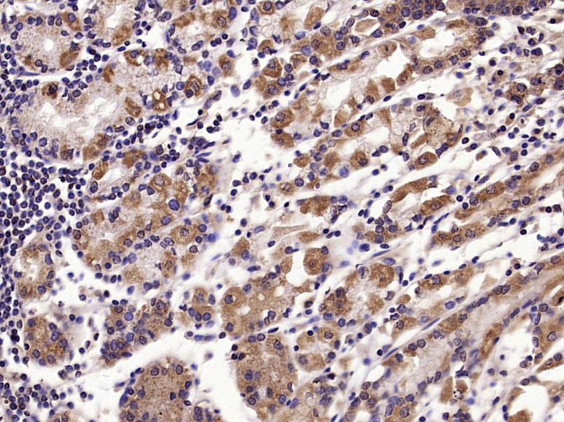 , ,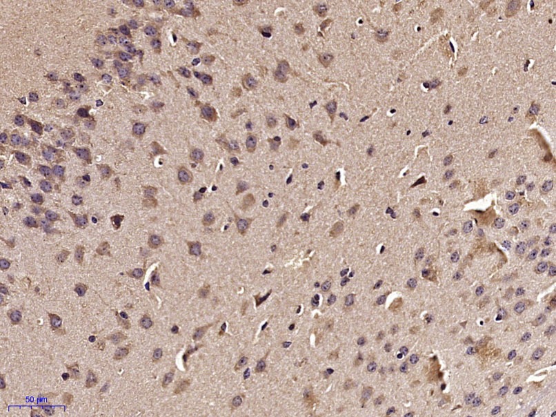 , ,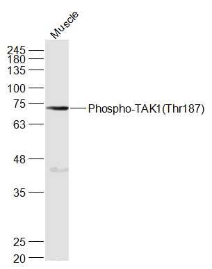 , ,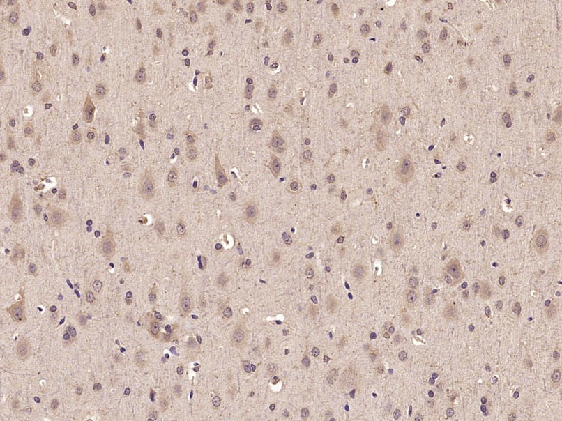 , ,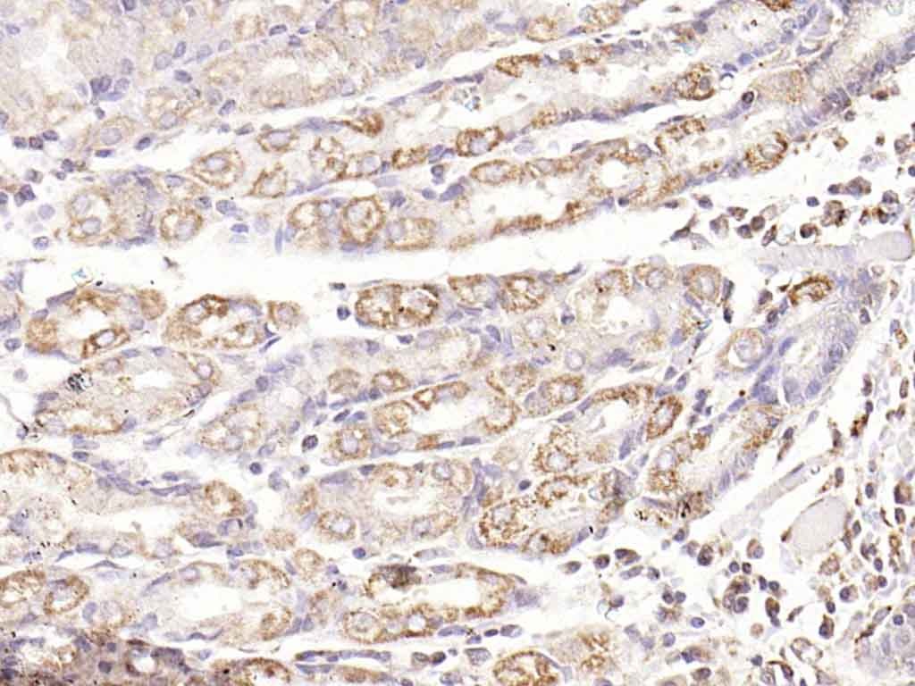 , ,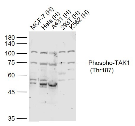 , ,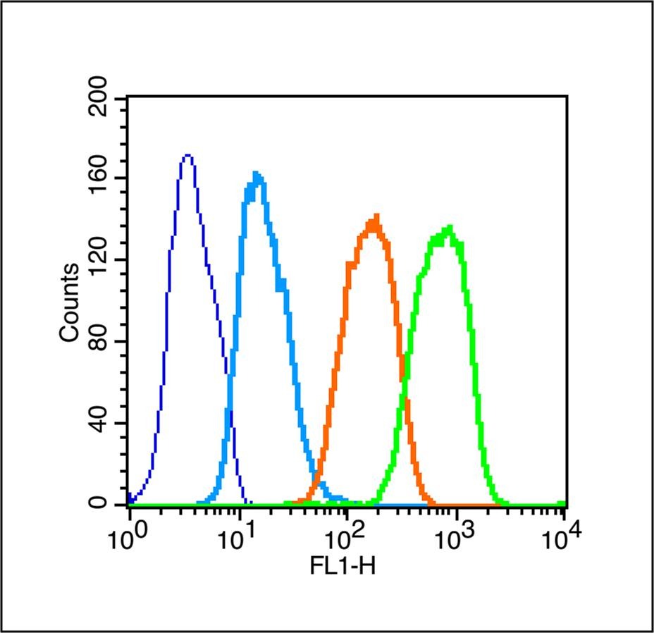 |
| Application | |
| Recommended Dose | WB: 1:500-2000; IHC-P: 1:100-500; IHC-Fr: 1:100-500; IF: 1:100-500; FCM: 1μg/Test |
| Antibody Type | Polyclonal |
| Host Species | Rabbit |
| Subcellular Localization | Cytoplasm. Cell membrane; Peripheral membrane protein; Cytoplasmic side. Note=Although the majority of MAP3K7/TAK1 is found in the cytosol, when complexed with TAB1/MAP3K7IP1 and TAB2/MAP3K7IP2, it is also localized at the cell membrane. |
| Tissue Specificity | Isoform 1A is the most abundant in ovary, skeletal muscle, spleen and blood mononuclear cells. Isoform 1B is highly expressed in brain, kidney and small intestine. Isoform 1C is the major form in prostate. Isoform 1D is the less abundant form. |
| Construction | Polyclonal Antibody |
| Purification | Protein A purified |
| Appearance | Liquid |
| Formulation | 0.01M TBS (pH7.4) with 1% BSA, 0.02% Proclin300 and 50% Glycerol. |
| Concentration | 1 mg/mL |
| Research Background | The protein encoded by this gene is a member of the serine/threonine protein kinase family. This kinase mediates the signaling transduction induced by TGF beta and morphogenetic protein (BMP), and controls a variety of cell functions including transcription regulation and apoptosis. In response to IL-1, this protein forms a kinase complex including TRAF6, MAP3K7P1/TAB1 and MAP3K7P2/TAB2; this complex is required for the activation of nuclear factor kappa B. This kinase can also activate MAPK8/JNK, MAP2K4/MKK4, and thus plays a role in the cell response to environmental stresses. Four alternatively spliced transcript variants encoding distinct isoforms have been reported. [provided by RefSeq, Jul 2008] |
| Immunogen | KLH conjugated Synthesised phosphopeptide: human TAK1 around the phosphorylation site of Thr187 |
| Antigen Species | Human |
| Gene Name | MAP3K7 |
| Gene ID | |
| Protein Name | Mitogen-activated protein kinase kinase kinase 7 |
| Uniprot ID | |
| Biology Area | Necroptosis,MAPK pathway,Inflammatory mediators,TLR Signaling,MAPK Pathway,Receptor Tyrosine Kinases,NFkB Pathway |
| Function | Component of a protein kinase signal transduction cascade. Mediator of TRAF6 and TGF-beta signal transduction. Activates IKBKB and MAPK8 in response to TRAF6 signaling. Stimulates NF-kappa-B activation and the p38 MAPK pathway. In osmotic stress signaling, plays a major role in the activation of MAPK8/JNK, but not that of NF-kappa-B. |
| Molecular Weight | Theoretical: 67 kDa. Actual: 72-75 kDa. |
| Stability & Storage | Store at -20°C or -80°C for 12 months. Avoid repeated freeze-thaw cycles. |
| Transport | Shipping with blue ice. |
| Size | Quantity | Unit Price | Amount | Operation |
|---|
 Hello! How can I help you today?
 Copyright © 2015-2026 TargetMol Chemicals Inc. All Rights Reserved.
