
Shopping Cart
Remove All Your shopping cart is currently empty

Synonyms: Smad2/3 (p-Thr8), Smad2/3 (p-T8), SMAD family member 3, SMAD family member 2, SMAD 3, SMAD 2, SMA and MAD related protein 3, Sma and Mad related protein 2, p-Smad2/3 (Thr8), p-Smad2/3 (T8), Mothers against DPP homolog 3, Mothers against DPP homolog 2, MADR2, MADH3, MADH2, Mad related protein 2, hSMAD3, hSMAD2, hMAD 3, hMAD 2
Anti-Phospho-Smad2/3
(Thr8) Polyclonal Antibody
| Pack Size | Price | USA Stock | Global Stock | Quantity |
|---|---|---|---|---|
| 50 µL | $222 | 7-10 days | 7-10 days | |
| 100 µL | $374 | 7-10 days | 7-10 days | |
| 200 µL | $527 | 7-10 days | 7-10 days |
| Description | Anti-Phospho-Smad2/3 (Thr8) Polyclonal Antibody is a Rabbit antibody targeting Phospho-Smad2/3 (Thr8). Anti-Phospho-Smad2/3 (Thr8) Polyclonal Antibody can be used in FCM,WB. |
| Synonyms | Smad2/3 (p-Thr8), Smad2/3 (p-T8), SMAD family member 3, SMAD family member 2, SMAD 3, SMAD 2, SMA and MAD related protein 3, Sma and Mad related protein 2, p-Smad2/3 (Thr8), p-Smad2/3 (T8), Mothers against DPP homolog 3, Mothers against DPP homolog 2, MADR2, MADH3, MADH2, Mad related protein 2, hSMAD3, hSMAD2, hMAD 3, hMAD 2 |
| Ig Type | IgG |
| Reactivity | Human,Mouse (predicted:Rat,Chicken,Dog,Pig,Cow,Horse) |
| Verified Activity | 1. Blank control: Hela. Primary Antibody (green line): Rabbit Anti-phospho-Smad2/Smad3 (Thr8) antibody (TMAB-01497) Dilution: 1 μg/10^6 cells; Isotype Control Antibody (orange line): Rabbit IgG. Secondary Antibody: Goat anti-rabbit IgG-AF647 Dilution: 1 μg/test. Protocol The cells were fixed with 4% PFA (10 min at room temperature) and then permeabilized with 90% ice-cold methanol for 20 min at-20°C. The cells were then incubated in 5% BSA to block non-specific protein-protein interactions for 30 min at room temperature. Cells stained with Primary Antibody for 30 min at room temperature. The secondary antibody used for 40 min at room temperature. 2. blank: A549 cells (blue line) isotype control: rabbit IgG (orange line) second antibody: goat anti-rabbit IgG (white blue line) primary antibody: rabbit Anti-phospho-Smad2/Smad3 (Thr8) (green line); contration: 3 μg/10^6 cells 3. Sample: HUVEC cell (human) Lysate at 30 μg Primary: Anti-p-Smad2/Smad3 (Thr8) (TMAB-01497) at 1/500 dilution Secondary: IRDye800CW Goat Anti-Rabbit IgG at 1/20000 dilution Predicted band size: 52 kDa Observed band size: 52 kDa 4. Sample: cerebrum (mouse) Lysate at 40 μg RSC96 cell (rat) Lysate at 30 μg hepG2 cell (human) Lysate at 30 μg Hcclm3 cell (human) Lysate at 30 μg Primary: Anti-p-Smad2/Smad3 (Thr8) (TMAB-01497) at 1/500 dilution Secondary: IRDye800CW Goat Anti-Rabbit IgG at 1/20000 dilution Predicted band size: 52 kDa Observed band size: 48,52 kDa 5. Sample: U251 cell (human) Lysate at 30 μg Primary: Anti-p-Smad2/Smad3 (Thr8) (TMAB-01497) at 1/500 dilution Secondary: IRDye800CW Goat Anti-Rabbit IgG at 1/20000 dilution Predicted band size: 52 kDa Observed band size: 52 kDa 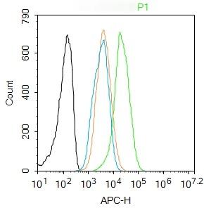 , ,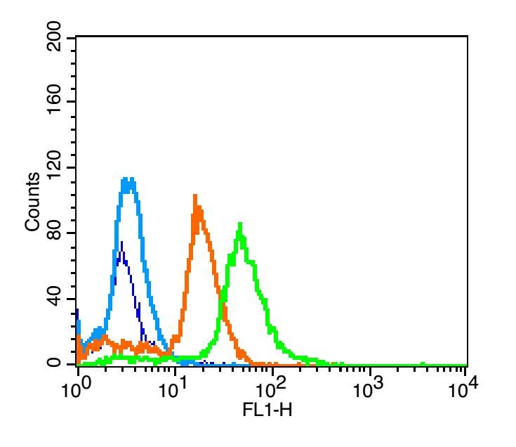 , ,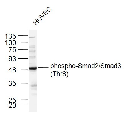 , ,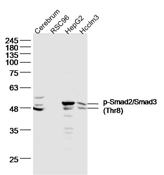 , ,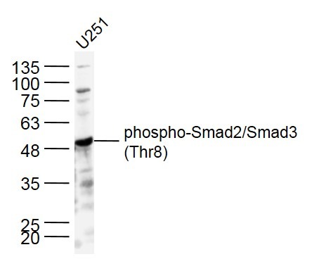 |
| Application | |
| Recommended Dose | WB: 1:500-2000; FCM: 1μg/Test |
| Antibody Type | Polyclonal |
| Host Species | Rabbit |
| Subcellular Localization | Cytoplasm. Nucleus. Note=Cytoplasmic and nuclear in the absence of TGF-beta. On TGF-beta stimulation, migrates to the nucleus when complexed with SMAD4. On dephosphorylation by phosphatase PPM1A, released from the SMAD2/SMAD4 complex, and exported out of the nucleus by interaction with RANBP1. |
| Tissue Specificity | Expressed at high levels in skeletal muscle, heart and placenta. |
| Construction | Polyclonal Antibody |
| Purification | Protein A purified |
| Appearance | Liquid |
| Formulation | 0.01M TBS (pH7.4) with 1% BSA, 0.02% Proclin300 and 50% Glycerol. |
| Concentration | 1 mg/mL |
| Research Background | The protein encoded by this gene belongs to the SMAD, a family of proteins similar to the gene products of the Drosophila gene 'mothers against decapentaplegic' (Mad) and the C. elegans gene Sma. SMAD proteins are signal transducers and transcriptional modulators that mediate multiple signaling pathways. This protein mediates the signal of the transforming growth factor (TGF)-beta, and thus regulates multiple cellular processes, such as cell proliferation, apoptosis, and differentiation. This protein is recruited to the TGF-beta receptors through its interaction with the SMAD anchor for receptor activation (SARA) protein. In response to TGF-beta signal, this protein is phosphorylated by the TGF-beta receptors. The phosphorylation induces the dissociation of this protein with SARA and the association with the family member SMAD4. The association with SMAD4 is important for the translocation of this protein into the nucleus, where it binds to target promoters and forms a transcription repressor complex with other cofactors. This protein can also be phosphorylated by activin type 1 receptor kinase, and mediates the signal from the activin. Alternatively spliced transcript variants have been observed for this gene. [provided by RefSeq, May 2012] |
| Immunogen | KLH conjugated synthesised phosphopeptide: human Smad2/Smad3 around the phosphorylation site of Thr8 |
| Antigen Species | Human |
| Gene Name | SMAD2 |
| Gene ID | |
| Protein Name | Mothers against decapentaplegic homolog 2 |
| Uniprot ID | |
| Biology Area | TGF,SMADs,SMADs,Cytoplasmic |
| Function | Receptor-regulated SMAD (R-SMAD) that is an intracellular signal transducer and transcriptional modulator activated by TGF-beta (transforming growth factor) and activin type 1 receptor kinases. Binds the TRE element in the promoter region of many genes that are regulated by TGF-beta and, on formation of the SMAD2/SMAD4 complex, activates transcription. May act as a tumor suppressor in colorectal carcinoma. Positively regulates PDPK1 kinase activity by stimulating its dissociation from the 14-3-3 protein YWHAQ which acts as a negative regulator. |
| Molecular Weight | Theoretical: 52 kDa. Actual: 48,52 kDa. |
| Stability & Storage | Store at -20°C or -80°C for 12 months. Avoid repeated freeze-thaw cycles. |
| Transport | Shipping with blue ice. |
| Size | Quantity | Unit Price | Amount | Operation |
|---|
 Hello! How can I help you today?
 Copyright © 2015-2026 TargetMol Chemicals Inc. All Rights Reserved.
