
Shopping Cart
Remove All Your shopping cart is currently empty


Anti-Pan Cytokeratin Polyclonal Antibody is a Rabbit antibody targeting Pan Cytokeratin. Anti-Pan Cytokeratin Polyclonal Antibody can be used in FCM,ICC/IF,IF,IHC-Fr,IHC-P,WB.
| Pack Size | Price | USA Warehouse | Global Warehouse | Quantity |
|---|---|---|---|---|
| 50 μL | $222 | 7-10 days | 7-10 days | |
| 100 μL | $373 | 7-10 days | 7-10 days | |
| 200 μL | $527 | 7-10 days | 7-10 days |
| Description | Anti-Pan Cytokeratin Polyclonal Antibody is a Rabbit antibody targeting Pan Cytokeratin. Anti-Pan Cytokeratin Polyclonal Antibody can be used in FCM,ICC/IF,IF,IHC-Fr,IHC-P,WB. |
| Synonyms | wide spectrum Cytokeratin, P-CK, pan-cytokeratin, pan-CK, Cytokeratins, CK-PAN, [cytokeratins 13, 14, 16, 17, 19, 24] |
| Ig Type | IgG |
| Reactivity | Human,Mouse,Rat,Cow (predicted:Chicken,Dog,Pig,Horse,Rabbit) |
| Verified Activity | 1. Tissue/cell: Rat brain tissue; 4% Paraformaldehyde-fixed and paraffin-embedded; Antigen retrieval: citrate buffer (0.01M, pH6.0), Boiling bathing for 15 min; Block endogenous peroxidase by 3% Hydrogen peroxide for 30 min; Blocking buffer (normal goat serum) at 37°C for 20 min; Incubation: Anti-Pan Cytokeratin Polyclonal Antibody, Unconjugated (TMAB-01329) 1:500, overnight at 4°C, followed by conjugation to the secondary antibody and DAb staining. 2. Tissue/cell: Human colon cancer; 4% Paraformaldehyde-fixed and paraffin-embedded; Antigen retrieval: citrate buffer (0.01M, pH6.0), Boiling bathing for 15 min; Block endogenous peroxidase by 3% Hydrogen peroxide for 30 min; Blocking buffer (normal goat serum) at 37°C for 20 min; Incubation: Anti-Pan Cytokeratin Polyclonal Antibody, Unconjugated (TMAB-01329) 1:500, overnight at 4°C, followed by conjugation to the secondary antibody and DAb staining. 3. Paraformaldehyde-fixed, paraffin embedded (mouse lung); Antigen retrieval by boiling in sodium citrate buffer (pH6) for 15 min; Block endogenous peroxidase by 3% hydrogen peroxide for 20 min; Blocking buffer (normal goat serum) at 37°C for 30 min; Antibody incubation with (Pan Cytokeratin) Polyclonal Antibody, Unconjugated (TMAB-01329) at 1:400 overnight at 4°C, followed by a conjugated secondary (Goat Anti-rabbit IgG/Bio) for 20 minutes at 37°C, followed by a conjugated streptavidin at[1:500] for 40 minutes and DAPI staining of the nucleus. 4. Sample: Lane 1: Hela (Human) Cell Lysate at 30 μg Lane 2: A549 (Human) Cell Lysate at 30 μg Lane 3: A673 (Human) Cell Lysate at 30 μg Primary: Anti-Pan Cytokeratin (TMAB-01329) at 1/1000 dilution Secondary: IRDye800CW Goat Anti-Rabbit IgG at 1/20000 dilution Predicted band size: 42-64 kDa Observed band size: 46,60 kDa 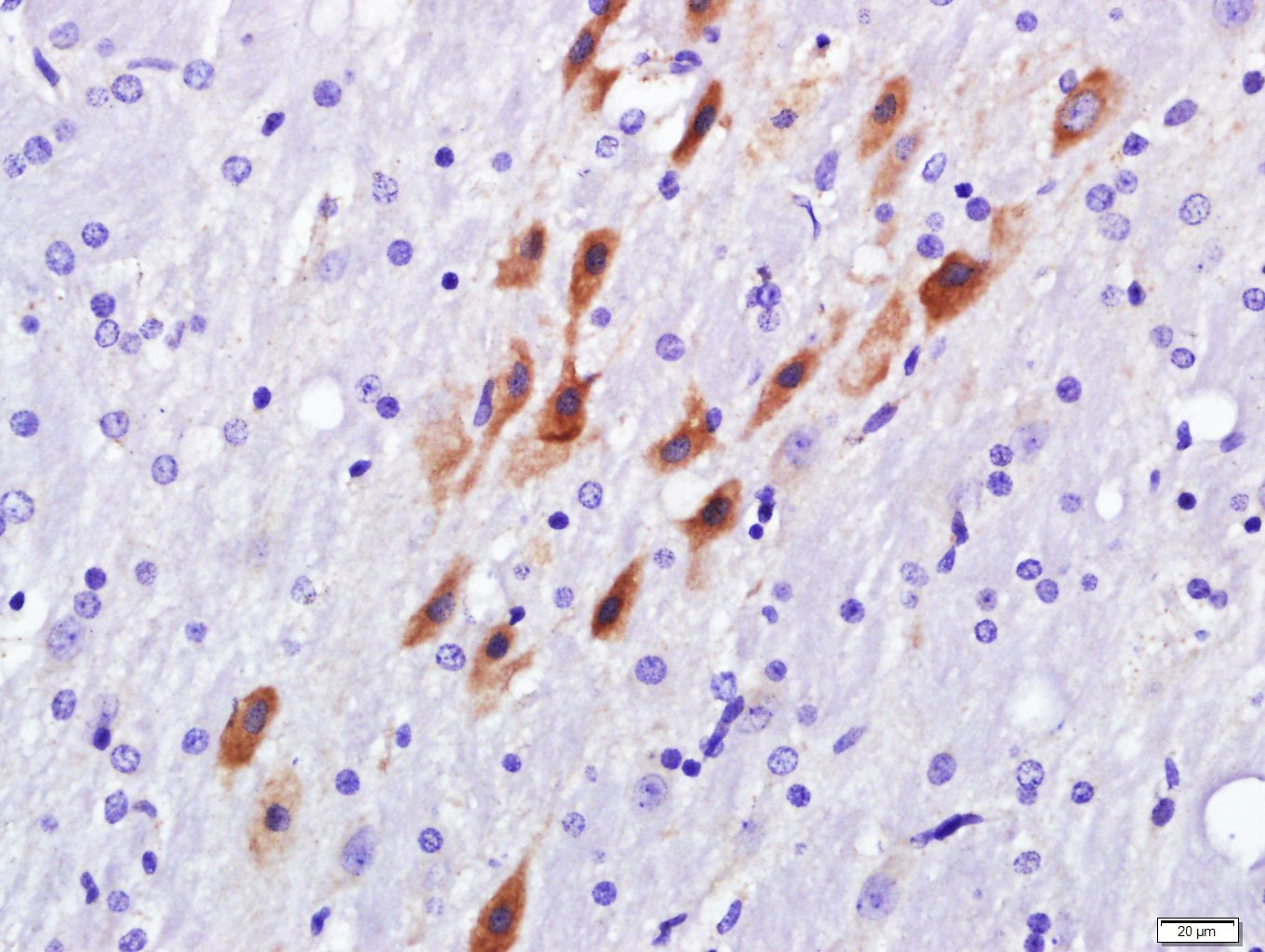 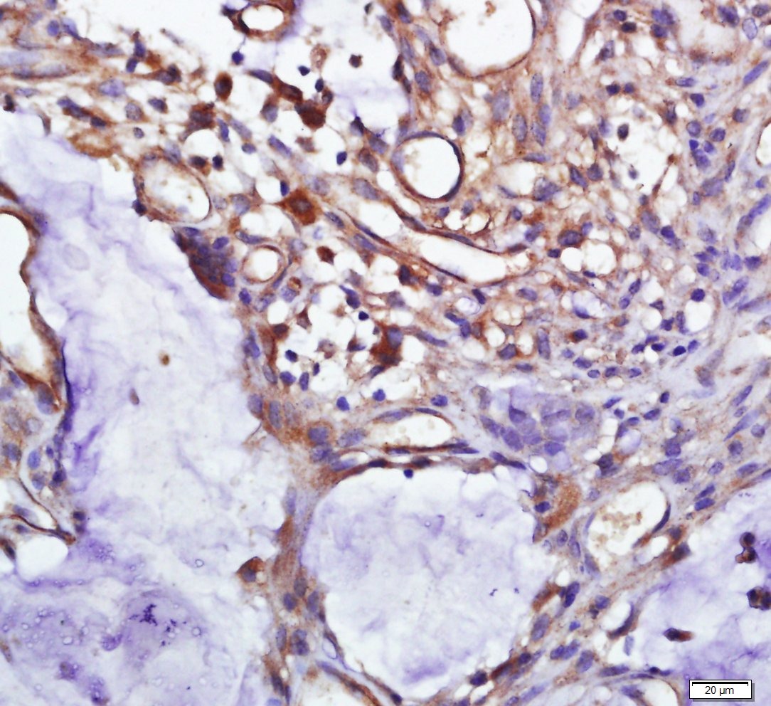 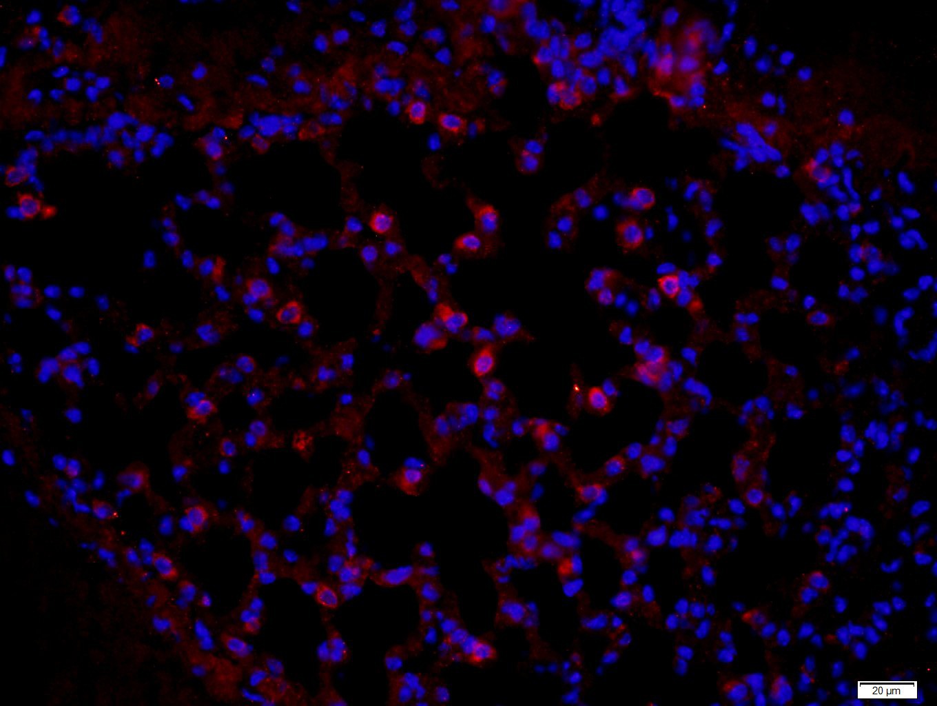 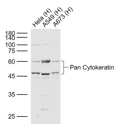 |
| Application | |
| Recommended Dose | WB: 1:500-2000; IHC-P: 1:100-2000; IHC-Fr: 1:100-500; ICC/IF: 1:100; IF: 1:100-500; FCM: 1μg /test |
| Antibody Type | Polyclonal |
| Host Species | Rabbit |
| Subcellular Localization | Cytoplasmic. |
| Tissue Specificity | epithelial cells |
| Construction | Polyclonal Antibody |
| Purification | Protein A purified |
| Appearance | Liquid |
| Formulation | 0.01M TBS (pH7.4) with 1% BSA, 0.02% Proclin300 and 50% Glycerol. |
| Concentration | 1 mg/mL |
| Research Background | Cytokeratins are proteins of keratin-containing intermediate filaments found in the intracytoplasmic cytoskeleton of epithelial tissue. The cytokeratins are encoded by a family encompassing 30 genes. Among them, 20 are epithelial genes and the remaining 10 are specific for trichocytes. In the cytoplasm, the keratin filaments conform a complex network which extends from the surface of the nucleus to the cell membrane. Numerous accessory proteins are involved in the genesis and maintenance of such structure. This association between the plasma membrane and the nuclear surface provides important implications for the organization of the cytoplasm and cellular communication mechanisms. Apart from the relatively static functions provided in terms of supporting the nucleus and providing tensile strength to the cell, the cytokeratin networks undergo rapid phosphate exchanges mediated depolymerization, with important implications in the more dynamic cellular processes such as mitosis and post-mitotic period, cell movement and differentiation. Cytokeratins interact with desmosomes and hemidesmosomes, thus collaborating to cell-cell adhesion and basal cell-underlying connective tissue connection. |
| Immunogen | KLH conjugated synthetic peptide: human cytokeratin 16 |
| Antigen Species | Human |
| Gene ID | |
| Uniprot ID | |
| Biology Area | Cytokeratins |
| Stability & Storage | Store at -20°C or -80°C for 12 months. Avoid repeated freeze-thaw cycles. |
| Transport | Shipping with blue ice. |

| Size | Quantity | Unit Price | Amount | Operation |
|---|
 Hello! How can I help you today?
 Copyright © 2015-2025 TargetMol Chemicals Inc. All Rights Reserved.

 For example, your dosage is 10 mg/kg Each animal weighs 20 g, and the dosage volume is 100 μL .