
Shopping Cart
Remove All Your shopping cart is currently empty

Synonyms:
Anti-MYLK Antibody
(1W61)
| Pack Size | Price | USA Stock | Global Stock | Quantity |
|---|---|---|---|---|
| 50 µL | $297 | 7-10 days | 7-10 days | |
| 100 µL | $497 | 7-10 days | 7-10 days |
| Description | Anti-MYLK Antibody (1W61) is a Rabbit antibody targeting MYLK. Anti-MYLK Antibody (1W61) can be used in FCM,ICC/IF,IHC,WB. |
| Ig Type | IgG |
| Clone | 1W61 |
| Reactivity | Human,Mouse,Rat |
| Verified Activity | 1. Western blot analysis of MYLK on human lung lysates using anti-MYLK antibody at 1/5,000 dilution. 2. Immunohistochemical analysis of paraffin-embedded rat smooth muscle tissue using anti-MYLK antibody. Counter stained with hematoxylin. 3. Immunohistochemical analysis of paraffin-embedded human colon cancer tissue using anti-MYLK antibody. Counter stained with hematoxylin. 4. Immunohistochemical analysis of paraffin-embedded mouse smooth muscle tissue using anti-MYLK antibody. Counter stained with hematoxylin. 5. ICC staining MYLK in Hela cells (green). The nuclear counter stain is DAPI (blue). Cells were fixed in paraformaldehyde, permeabilised with 0.25% Triton X100/PBS. 6. ICC staining MYLK in L6 cells (green). The nuclear counter stain is DAPI (blue). Cells were fixed in paraformaldehyde, permeabilised with 0.25% Triton X100/PBS. 7. ICC staining MYLK in NIH/3T3 cells (green). The nuclear counter stain is DAPI (blue). Cells were fixed in paraformaldehyde, permeabilised with 0.25% Triton X100/PBS. 8. ICC staining MYLK in SH-SY-5Y cells (green). The nuclear counter stain is DAPI (blue). Cells were fixed in paraformaldehyde, permeabilised with 0.25% Triton X100/PBS. 9. Flow cytometric analysis of SH-SY-5Y cells with MYLK antibody at 1/50 dilution (red) compared with an unlabelled control (cells without incubation with primary antibody; black). Alexa Fluor 488-conjugated goat anti rabbit IgG was used as the secondary antibody. 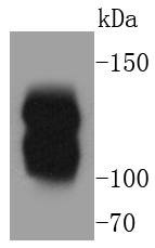 , ,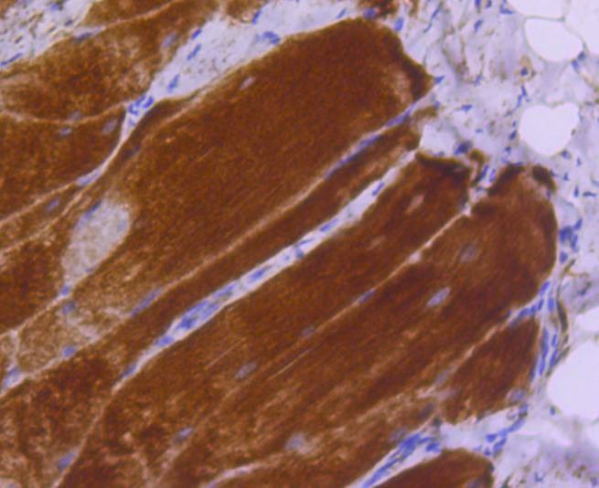 , ,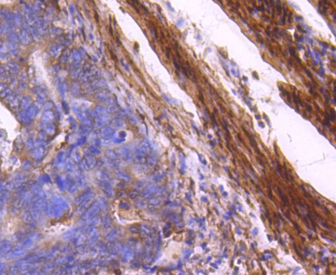 , ,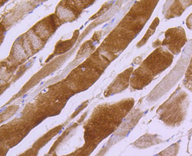 , ,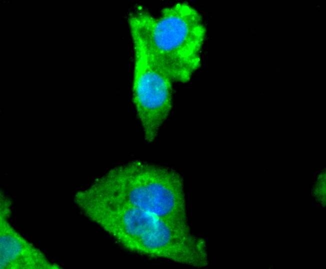 , ,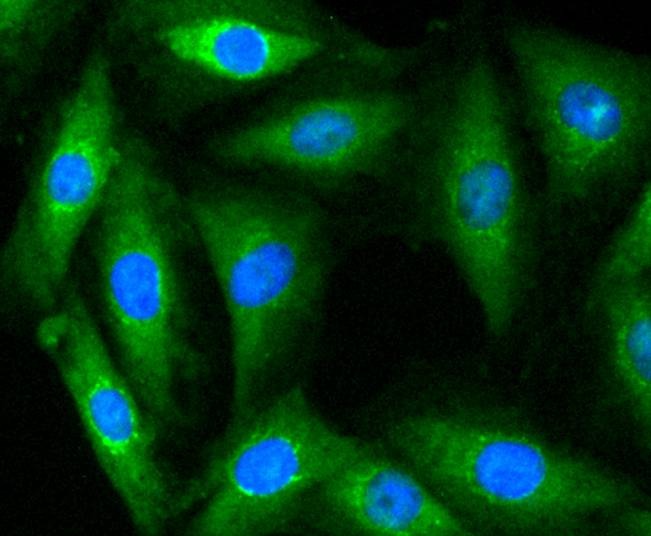 , ,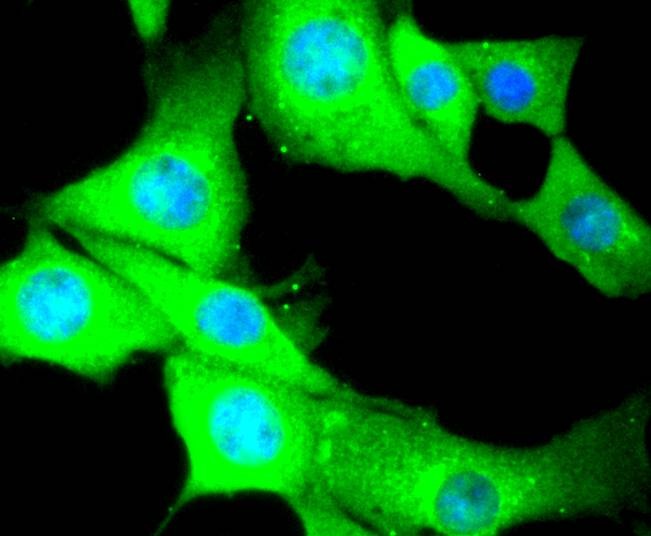 , ,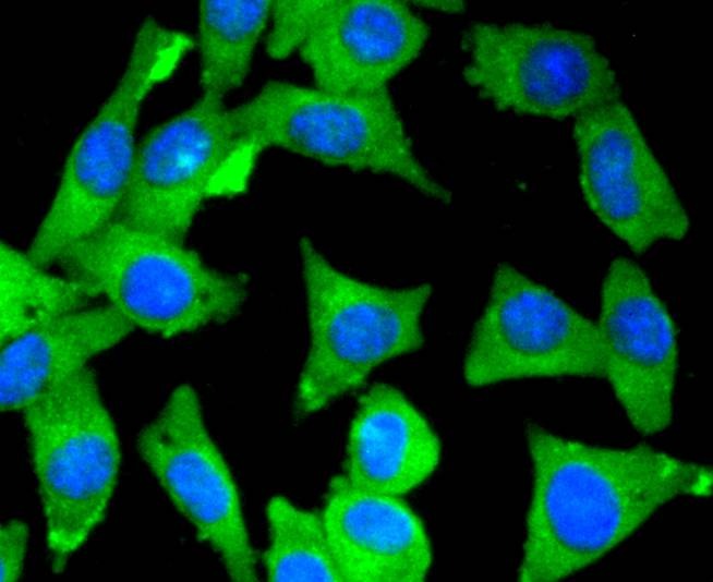 , ,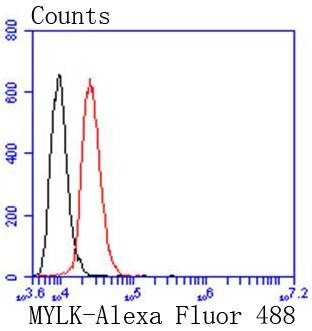 |
| Application | |
| Recommended Dose | WB: 1:2000-10000; IHC: 1:100-500; ICC/IF: 1:100-500; FCM: 1:50-100 |
| Antibody Type | Monoclonal |
| Host Species | Rabbit |
| Construction | Recombinant Antibody |
| Purification | ProA affinity purified |
| Appearance | Liquid |
| Formulation | 1*TBS (pH7.4), 1%BSA, 40%Glycerol. Preservative: 0.05% Sodium Azide. |
| Research Background | MLCK, a member of the Ser/Thr protein kinase family, is a calcium/calmodulin-dependent enzyme responsible for smooth muscle contraction via phosphorylation of a specific serine in the N-terminus of myosin light chains (MLC), an event that facilitates myosin interaction with actin filaments. It is a central determinant in the development of vascular permeability and tissue edema formation. In the nervous system it has been shown to control the growth initiation of astrocytic processes in culture and to participate in transmitter release at synapses formed between cultured sympathetic ganglion cells. MLCK acts as a critical participant in signaling sequences that result in fibroblast apoptosis. Smooth muscle and non-muscle isozymes are expressed in a wide variety of adult and fetal tissues and in cultured endothelium with qualitative expression appearing to be neither tissue- nor development-specific. Non-muscle isoform 2 is the dominant splice variant expressed in various tissues. The Telokin isoform, which binds calmodulin, has been found in a wide variety of adult and fetal tissues. MLCK is probably down-regulated by phosphorylation. The protein contains 1 fibronectin type III domain and 9 immunoglobulin-like C2-type domains. |
| Conjucates | Unconjugated |
| Immunogen | Recombinant Protein |
| Uniprot ID |
| Molecular Weight | Theoretical: 210 kDa. |
| Stability & Storage | Store at -20°C or -80°C for 12 months. Avoid repeated freeze-thaw cycles. |
| Transport | Shipping with blue ice. |
| Size | Quantity | Unit Price | Amount | Operation |
|---|
 Hello! How can I help you today?
 Copyright © 2015-2026 TargetMol Chemicals Inc. All Rights Reserved.
