
Shopping Cart
Remove All Your shopping cart is currently empty

Synonyms: Microtubule-associated proteins 1A/1B light chain 3A, Microtubule-associated protein 1 lig, MAP1A/MAP1B light chain 3 A, MAP1A/MAP1B LC3 A, MAP1 light chain 3-like protein 2, Autophagy-related ubiquitin-like modifier LC3 A, Autophagy-related protein LC3 A
Anti-LC3A Antibody
(4U976)
| Pack Size | Price | USA Stock | Global Stock | Quantity |
|---|---|---|---|---|
| 50 µL | $220 | 7-10 days | 7-10 days | |
| 100 µL | $373 | 7-10 days | 7-10 days | |
| 200 µL | $529 | 7-10 days | 7-10 days |
| Description | Anti-LC3A Antibody (4U976) is a Mouse antibody targeting ADAR1. Anti-LC3A Antibody (4U976) can be used in IF,IHC-Fr,IHC-P,WB. |
| Synonyms | Microtubule-associated proteins 1A/1B light chain 3A, Microtubule-associated protein 1 lig, MAP1A/MAP1B light chain 3 A, MAP1A/MAP1B LC3 A, MAP1 light chain 3-like protein 2, Autophagy-related ubiquitin-like modifier LC3 A, Autophagy-related protein LC3 A |
| Ig Type | IgG |
| Clone | 4U976 |
| Reactivity | Human,Mouse,Rat |
| Verified Activity | 1. Paraformaldehyde-fixed, paraffin embedded (Mouse brain); Antigen retrieval by boiling in sodium citrate buffer (pH6.0) for 15 min; Block endogenous peroxidase by 3% hydrogen peroxide for 20 min; Blocking buffer (normal goat serum) at 37°C for 30 min; Antibody incubation with (LC3A) Monoclonal Antibody, Unconjugated (TMAB-01098) at 1:400 overnight at 4°C, followed by a conjugated secondary for 20 min and DAB staining.
2. Paraformaldehyde-fixed, paraffin embedded (Rat brain); Antigen retrieval by boiling in sodium citrate buffer (pH6.0) for 15 min; Block endogenous peroxidase by 3% hydrogen peroxide for 20 min; Blocking buffer (normal goat serum) at 37°C for 30 min; Antibody incubation with (LC3A) Monoclonal Antibody, Unconjugated (TMAB-01098) at 1:400 overnight at 4°C, followed by a conjugated secondary for 20 min and DAB staining. 3. Paraformaldehyde-fixed, paraffin embedded (Mouse brain); Antigen retrieval by boiling in sodium citrate buffer (pH6.0) for 15 min; Block endogenous peroxidase by 3% hydrogen peroxide for 20 min; Blocking buffer (normal goat serum) at 37°C for 30 min; Antibody incubation with (LC3A) Monoclonal Antibody, Unconjugated (TMAB-01098) at 1:400 overnight at 4°C, followed by a conjugated Goat Anti-Mouse IgG antibody for 90 minutes, and DAPI for nucleus staining. 4. Paraformaldehyde-fixed, paraffin embedded (Rat brain); Antigen retrieval by boiling in sodium citrate buffer (pH6.0) for 15 min; Block endogenous peroxidase by 3% hydrogen peroxide for 20 min; Blocking buffer (normal goat serum) at 37°C for 30 min; Antibody incubation with (LC3A) Monoclonal Antibody, Unconjugated (TMAB-01098) at 1:400 overnight at 4°C, followed by a conjugated Goat Anti-Mouse IgG antibody for 90 minutes, and DAPI for nucleus staining. 5. Paraformaldehyde-fixed, paraffin embedded (rat cerebellum); Antigen retrieval by boiling in sodium citrate buffer (pH6.0) for 15 min; Block endogenous peroxidase by 3% hydrogen peroxide for 20 min; Blocking buffer (normal goat serum) at 37°C for 30 min; Incubation with (LC3A) Monoclonal Antibody, Unconjugated (TMAB-01098) at 1:200 overnight at 4°C, followed by operating according to SP Kit (Mouse) instructionsand DAB staining. 6. Paraformaldehyde-fixed, paraffin embedded (human cerebellum); Antigen retrieval by boiling in sodium citrate buffer (pH6.0) for 15 min; Block endogenous peroxidase by 3% hydrogen peroxide for 20 min; Blocking buffer (normal goat serum) at 37°C for 30 min; Incubation with (LC3A) Monoclonal Antibody, Unconjugated (TMAB-01098) at 1:200 overnight at 4°C, followed by operating according to SP Kit (Mouse) instructionsand DAB staining. 7. Paraformaldehyde-fixed, paraffin embedded (mouse cerebellum); Antigen retrieval by boiling in sodium citrate buffer (pH6.0) for 15 min; Block endogenous peroxidase by 3% hydrogen peroxide for 20 min; Blocking buffer (normal goat serum) at 37°C for 30 min; Incubation with (LC3A) Monoclonal Antibody, Unconjugated (TMAB-01098) at 1:200 overnight at 4°C, followed by operating according to SP Kit (Mouse) instructionsand DAB staining. 8. Paraformaldehyde-fixed, paraffin embedded (human brain); Antigen retrieval by boiling in sodium citrate buffer (pH6.0) for 15 min; Block endogenous peroxidase by 3% hydrogen peroxide for 20 min; Blocking buffer (normal goat serum) at 37°C for 30 min; Incubation with (LC3A) Monoclonal Antibody, Unconjugated (TMAB-01098) at 1:200 overnight at 4°C, followed by operating according to SP Kit (Mouse) instructionsand DAB staining. 9. Sample: Lane 1: Mouse Cerebrum tissue lysates Lane 2: Rat Cerebrum tissue lysates Lane 3: Rat Cerebellum tissue lysates Primary: Anti-LC3A (TMAB-01098) at 1/1000 dilution Secondary: IRDye800CW Goat Anti-Mouse IgG at 1/20000 dilution Predicted band size: 14/16 kDa Observed band size: 15 kDa 10. SH-SY5Y cell; 4% Paraformaldehyde-fixed; Triton X-100 at room temperature for 20 min; Blocking buffer (normal goat serum) at 37°C for 20 min; Antibody incubation with (LC3A) polyclonal Antibody, Unconjugated (TMAB-01098) 1:25, 90 minutes at 37°C; followed by a conjugated Goat Anti-Mouse IgG antibody at 37°C for 90 minutes, DAPI (blue) was used to stain the cell nucleus. 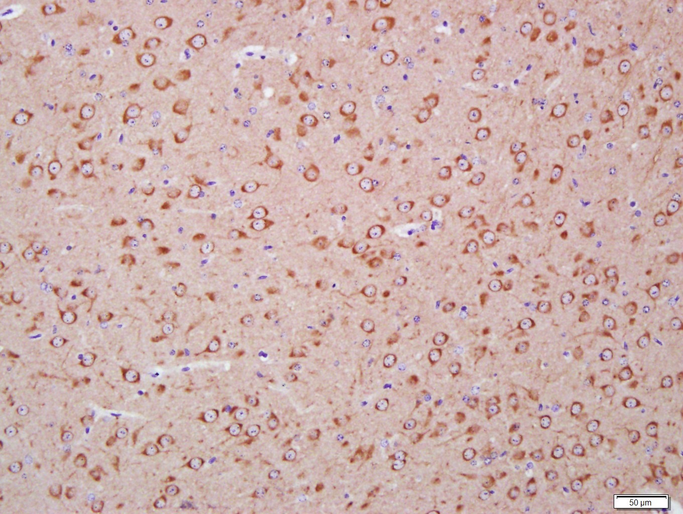 , ,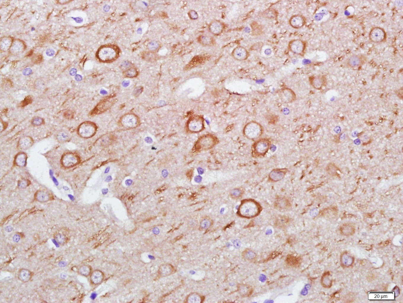 , ,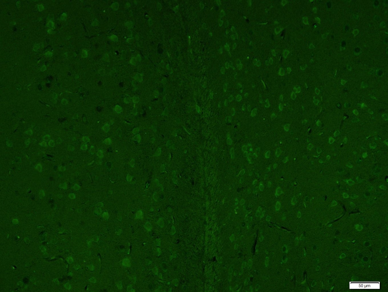 , ,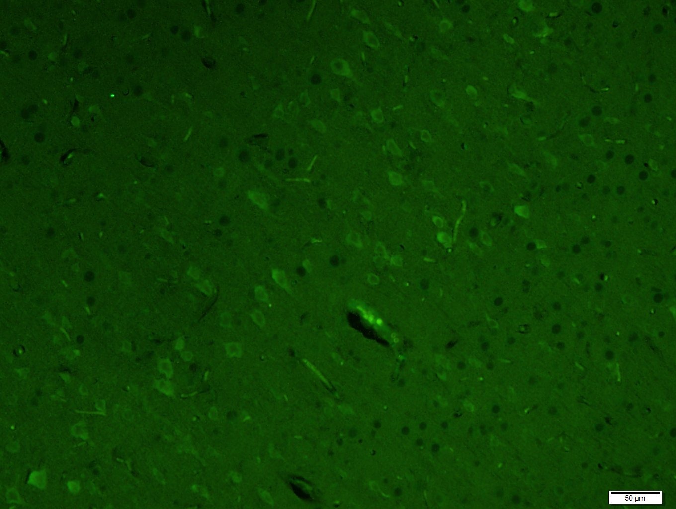 , ,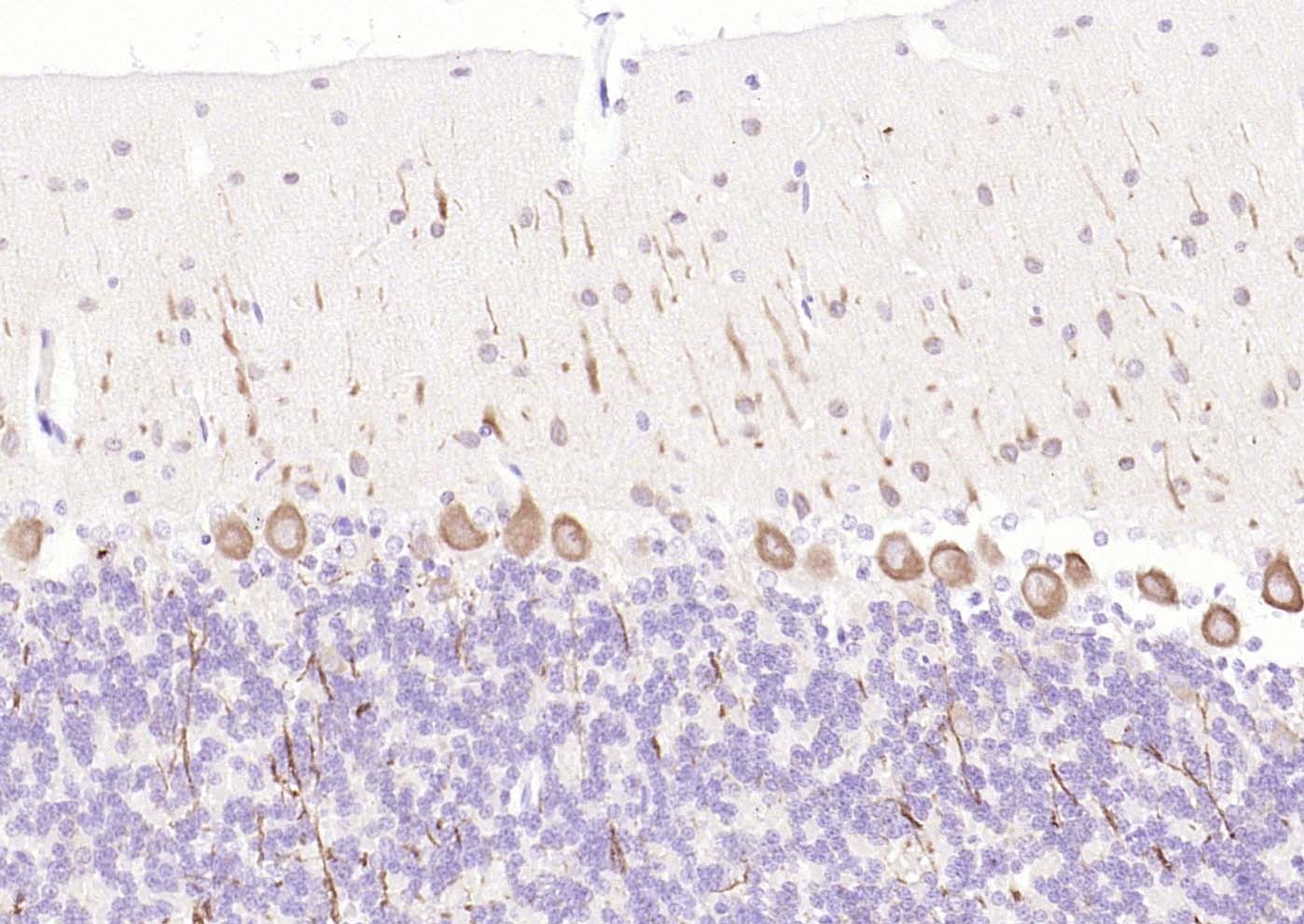 , ,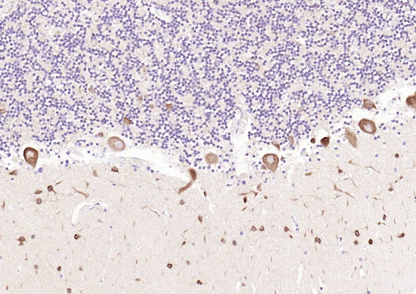 , ,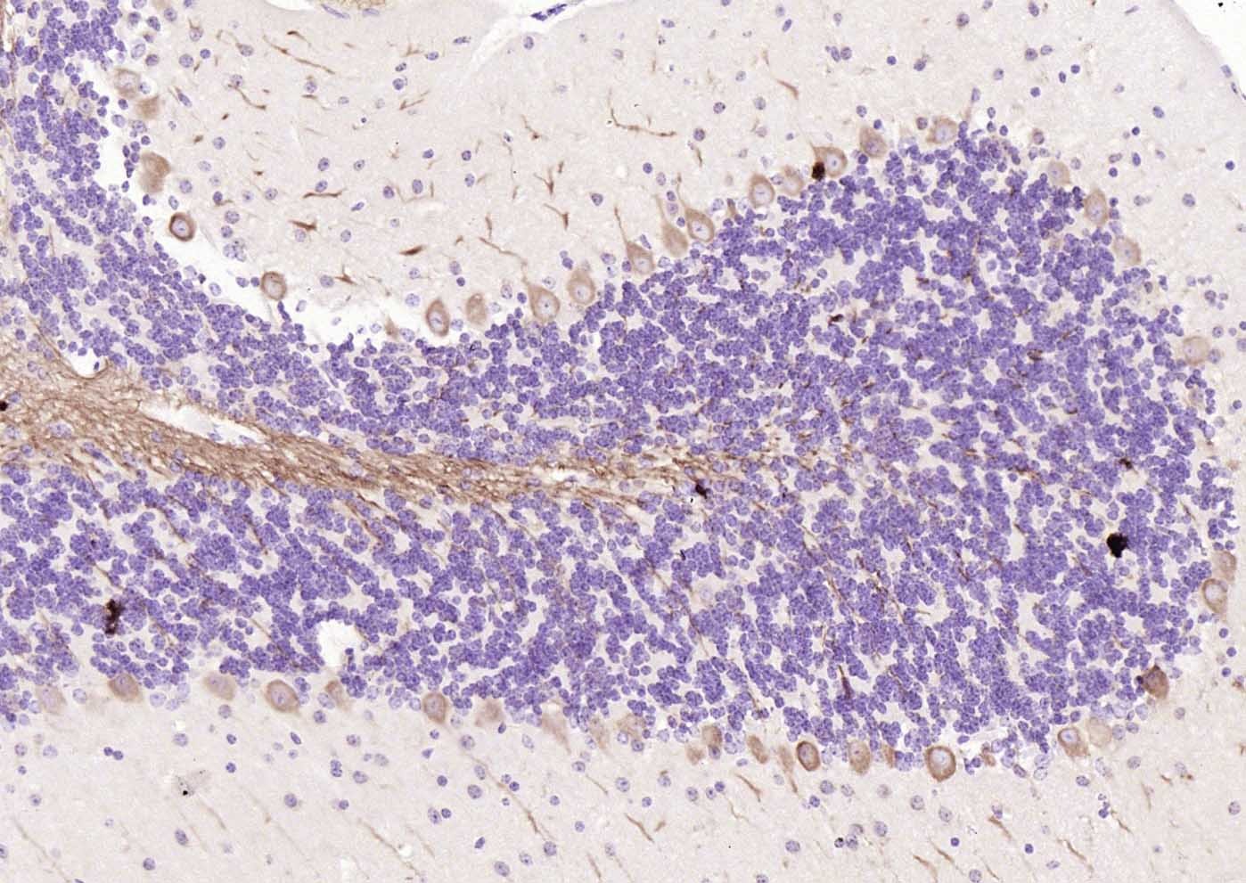 , ,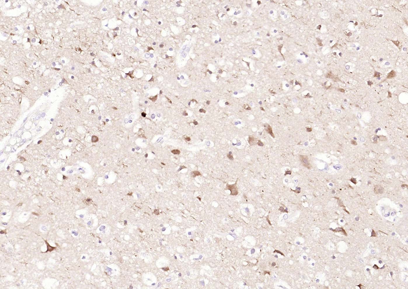 , ,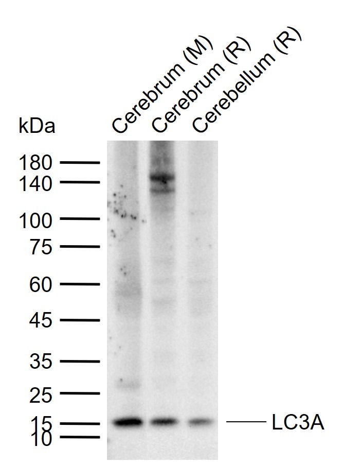 , ,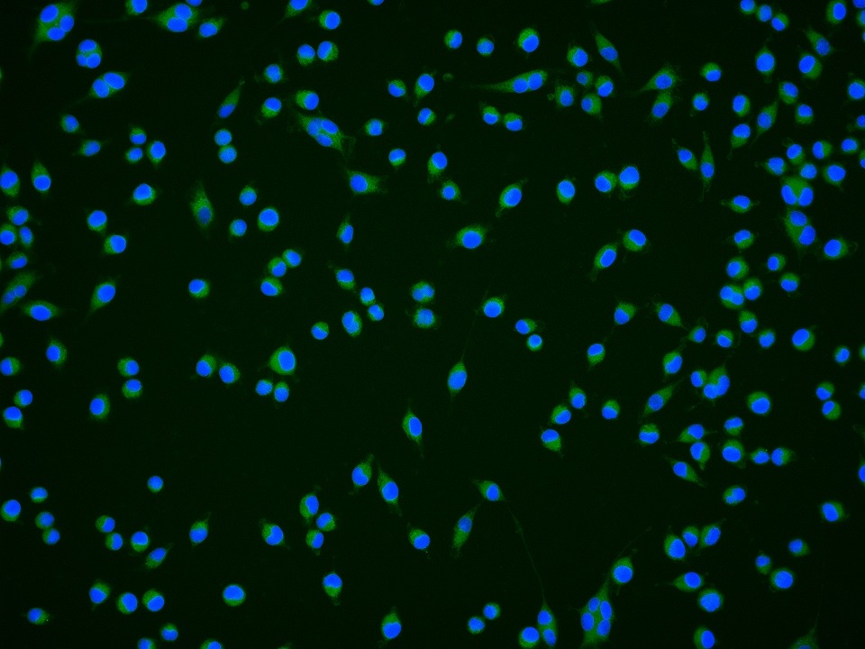 |
| Application | |
| Recommended Dose | IF=1:100-500; IHC-Fr=1:100-500; IHC-P=1:100-500; WB=1:500-1000 |
| Antibody Type | Monoclonal |
| Host Species | Mouse |
| Subcellular Localization | Cytoplasmic. Endomembrane system; Lipid-anchor. Cytoplasmic vesicle, autophagosome membrane; Lipid-anchor. Note: LC3B binds to the autophagic membranes. |
| Tissue Specificity | Most abundant in heart, brain, liver, skeletal muscle and testis but absent in thymus and peripheral blood leukocytes. |
| Construction | Hybridoma Monoclonal Antibody |
| Purification | Protein G purified |
| Appearance | Liquid |
| Formulation | 0.01M TBS (pH7.4) with 1% BSA, 0.02% Proclin300 and 50% Glycerol. |
| Concentration | 1 mg/mL |
| Research Background | A major contributor to cellular homeostasis is the ability of the cell to strike a balance between the formation and degradation/removal of its cellular components. This process of internal cellular turn-over is called autophagy (self-eating), and is facilitated by a pathway of around 16 interacting proteins in the human. LC3, a ubiquitin-like modifier protein, is the human homolog of yeast Apg8 and is involved in the formation of autophagosomal vacuoles, called autophagosomes. LC3 is expressed as 3 splice variants (LC3A, LC3B and LC3C), which exhibit different tissue distributions and are processed into cytosolic and autophagosomal membrane-bound forms, termed LC3-I and LC3-II, respectively. A disruption to the autophagic process is now associated with the progression of several cancers, neurodegenerative disorders and cardiac pathologies, where LC3 is widely employed as a marker for autophagy. |
| Immunogen | KLH conjugated synthetic peptide: human LC3A |
| Antigen Species | Human |
| Gene Name | MAP1LC3A |
| Gene ID | |
| Protein Name | Microtubule-associated proteins 1A/1B light chain 3A |
| Uniprot ID | |
| Biology Area | APG gene products,Signal Transduction,Autophagy,APG gene products,Autophagy and mitophagy,APG gene products,Neurogenesis,MAP |
| Function | Probably involved in formation of autophagosomal vacuoles (autophagosomes). |
| Molecular Weight | Theoretical: 14/16 kDa. Actual: 15 kDa. |
| Stability & Storage | Store at -20°C or -80°C for 12 months. Avoid repeated freeze-thaw cycles. |
| Transport | Shipping with blue ice. |
| Size | Quantity | Unit Price | Amount | Operation |
|---|
 Hello! How can I help you today?
 Copyright © 2015-2026 TargetMol Chemicals Inc. All Rights Reserved.
