
Shopping Cart
Remove All Your shopping cart is currently empty

Synonyms: TROP-1, TROP1, TACSTD1, MK-1, MIC18, M4S1, KSA, KS1/4, HNPCC8, ESA, epithelial cell adhesion molecule, EGP40, EGP314, EGP-2, DIAR5
Anti-EpCAM Polyclonal Antibody 2
| Pack Size | Price | USA Stock | Global Stock | Quantity |
|---|---|---|---|---|
| 50 µL | $220 | 7-10 days | 7-10 days | |
| 100 µL | $374 | 7-10 days | 7-10 days | |
| 200 µL | $527 | 7-10 days | 7-10 days |
| Description | Anti-EpCAM Polyclonal Antibody 2 is a Rabbit antibody targeting EpCAM. Anti-EpCAM Polyclonal Antibody 2 can be used in FCM,IF,IHC-Fr,IHC-P,WB. |
| Synonyms | TROP-1, TROP1, TACSTD1, MK-1, MIC18, M4S1, KSA, KS1/4, HNPCC8, ESA, epithelial cell adhesion molecule, EGP40, EGP314, EGP-2, DIAR5 |
| Ig Type | IgG |
| Reactivity | Human,Mouse,Rat |
| Verified Activity | 1. Paraformaldehyde-fixed, paraffin embedded (Human stomach cancer); Antigen retrieval by boiling in sodium citrate buffer (pH6.0) for 15 min; Block endogenous peroxidase by 3% hydrogen peroxide for 20 min; Blocking buffer (normal goat serum) at 37°C for 30 min; Antibody incubation with (EpCAM) Polyclonal Antibody, Unconjugated (TMAB-00623) at 1:400 overnight at 4°C, followed by operating according to SP Kit (Rabbit) instructionsand DAB staining. 2. Paraformaldehyde-fixed, paraffin embedded (rat colon); Antigen retrieval by boiling in sodium citrate buffer (pH6.0) for 15 min; Block endogenous peroxidase by 3% hydrogen peroxide for 20 min; Blocking buffer (normal goat serum) at 37°C for 30 min; Antibody incubation with (EpCAM) Polyclonal Antibody, Unconjugated (TMAB-00623) at 1:200 overnight at 4°C, followed by operating according to SP Kit (Rabbit) instructionsand DAB staining. 3. Sample: Lane 1: A431 (Human) Cell Lysate at 30 μg Lane 2: MCF-7 (Human) Cell Lysate at 30 μg Lane 3: HepG2 (Human) Cell Lysate at 30 μg Lane 4: K562 (Human) Cell Lysate at 30 μg Primary: Anti-EpCAM (TMAB-00623) at 1/1000 dilution Secondary: IRDye800CW Goat Anti-Rabbit IgG at 1/20000 dilution Predicted band size: 35 kDa Observed band size: 38 kDa 4. Paraformaldehyde-fixed, paraffin embedded (human colon); Antigen retrieval by boiling in sodium citrate buffer (pH6.0) for 15 min; Block endogenous peroxidase by 3% hydrogen peroxide for 20 min; Blocking buffer (normal goat serum) at 37°C for 30 min; Antibody incubation with (EpCAM) Polyclonal Antibody, Unconjugated (TMAB-00623) at 1:200 overnight at 4°C, followed by operating according to SP Kit (Rabbit) instructionsand DAB staining. 5. Blank control: FHC. Primary Antibody (green line): Rabbit Anti-EpCAM antibody (TMAB-00623) Dilution: 1 μg/Test; Secondary Antibody: Goat anti-rabbit IgG-FITC Dilution: 0.5 μg/Test. Protocol The cells were incubated in 5% BSA to block non-specific protein-protein interactions for 30 min at room temperature. Cells stained with Primary Antibody for 30 min at room temperature. The secondary antibody used for 40 min at room temperature. 6. Paraformaldehyde-fixed, paraffin embedded (rat kidney); Antigen retrieval by boiling in sodium citrate buffer (pH6.0) for 15 min; Block endogenous peroxidase by 3% hydrogen peroxide for 20 min; Blocking buffer (normal goat serum) at 37°C for 30 min; Antibody incubation with (EpCAM) Polyclonal Antibody, Unconjugated (TMAB-00623) at 1:200 overnight at 4°C, followed by operating according to SP Kit (Rabbit) instructionsand DAB staining. 7. Paraformaldehyde-fixed, paraffin embedded (rat uterus); Antigen retrieval by boiling in sodium citrate buffer (pH6.0) for 15 min; Block endogenous peroxidase by 3% hydrogen peroxide for 20 min; Blocking buffer (normal goat serum) at 37°C for 30 min; Antibody incubation with (EpCAM) Polyclonal Antibody, Unconjugated (TMAB-00623) at 1:200 overnight at 4°C, followed by operating according to SP Kit (Rabbit) instructionsand DAB staining. 8. Paraformaldehyde-fixed, paraffin embedded (Human duodenum); Antigen retrieval by boiling in sodium citrate buffer (pH6.0) for 15 min; Block endogenous peroxidase by 3% hydrogen peroxide for 20 min; Blocking buffer (normal goat serum) at 37°C for 30 min; Antibody incubation with (EpCAM) Polyclonal Antibody, Unconjugated (TMAB-00623) at 1:200 overnight at 4°C, followed by operating according to SP Kit (Rabbit) instructionsand DAB staining. 9. Paraformaldehyde-fixed, paraffin embedded (human colon carcinoma); Antigen retrieval by boiling in sodium citrate buffer (pH6.0) for 15 min; Block endogenous peroxidase by 3% hydrogen peroxide for 20 min; Blocking buffer (normal goat serum) at 37°C for 30 min; Antibody incubation with (EpCAM) Polyclonal Antibody, Unconjugated (TMAB-00623) at 1:200 overnight at 4°C, followed by operating according to SP Kit (Rabbit) instructionsand DAB staining. 10. Paraformaldehyde-fixed, paraffin embedded (mouse prostate); Antigen retrieval by boiling in sodium citrate buffer (pH6.0) for 15 min; Block endogenous peroxidase by 3% hydrogen peroxide for 20 min; Blocking buffer (normal goat serum) at 37°C for 30 min; Antibody incubation with (EpCAM) Polyclonal Antibody, Unconjugated (TMAB-00623) at 1:200 overnight at 4°C, followed by operating according to SP Kit (Rabbit) instructionsand DAB staining. 11. Paraformaldehyde-fixed, paraffin embedded (mouse uterus); Antigen retrieval by boiling in sodium citrate buffer (pH6.0) for 15 min; Block endogenous peroxidase by 3% hydrogen peroxide for 20 min; Blocking buffer (normal goat serum) at 37°C for 30 min; Antibody incubation with (EpCAM) Polyclonal Antibody, Unconjugated (TMAB-00623) at 1:200 overnight at 4°C, followed by operating according to SP Kit (Rabbit) instructionsand DAB staining. 12. Paraformaldehyde-fixed, paraffin embedded (rat colon); Antigen retrieval by boiling in sodium citrate buffer (pH6.0) for 15 min; Block endogenous peroxidase by 3% hydrogen peroxide for 20 min; Blocking buffer (normal goat serum) at 37°C for 30 min; Antibody incubation with (EpCAM) Polyclonal Antibody, Unconjugated (TMAB-00623) at 1:200 overnight at 4°C, followed by operating according to SP Kit (Rabbit) instructionsand DAB staining. 13. Paraformaldehyde-fixed, paraffin embedded (rat small intestine); Antigen retrieval by boiling in sodium citrate buffer (pH6.0) for 15 min; Block endogenous peroxidase by 3% hydrogen peroxide for 20 min; Blocking buffer (normal goat serum) at 37°C for 30 min; Antibody incubation with (EpCAM) Polyclonal Antibody, Unconjugated (TMAB-00623) at 1:200 overnight at 4°C, followed by operating according to SP Kit (Rabbit) instructionsand DAB staining. 14. Blank control: K562. Primary Antibody (green line): Rabbit Anti-EpCAM antibody (TMAB-00623) Dilution: 1 μg/10^6 cells; Isotype Control Antibody (orange line): Rabbit IgG. Secondary Antibody: Goat anti-rabbit IgG-FITC Dilution: 0.5 μg/test. Protocol The cells were incubated in 5% BSA to block non-specific protein-protein interactions for 30 min at room temperature. Cells stained with Primary Antibody for 30 min at room temperature. The secondary antibody used for 40 min at room temperature. 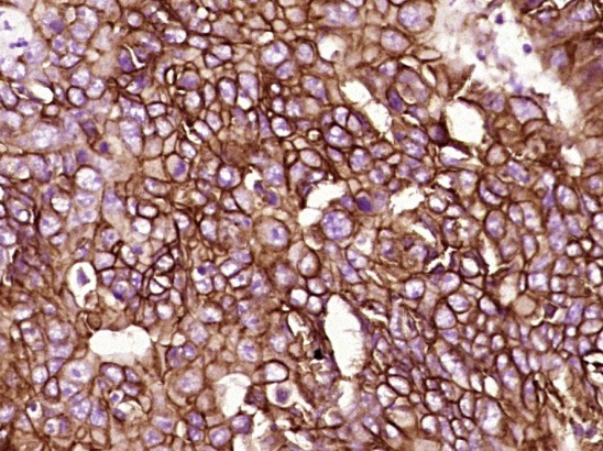 , ,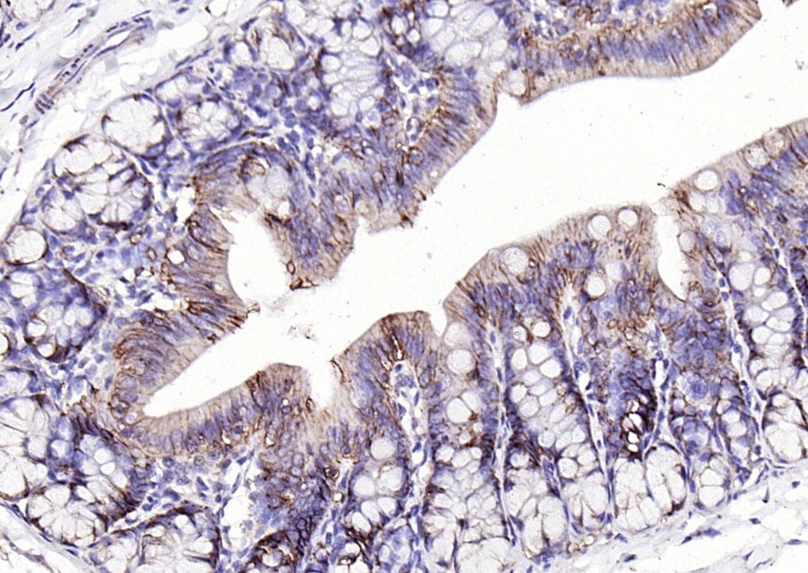 , ,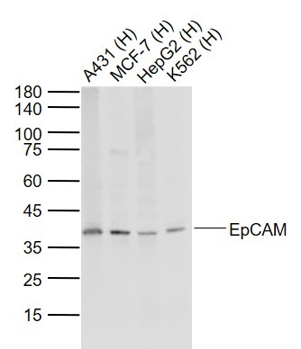 , ,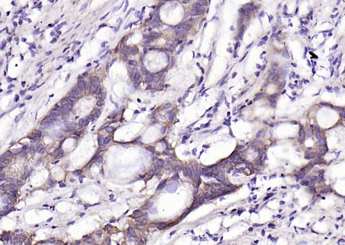 , ,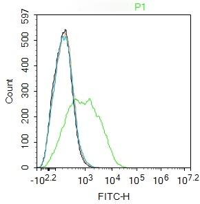 , ,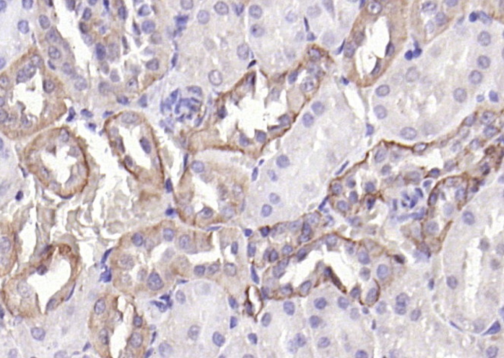 , ,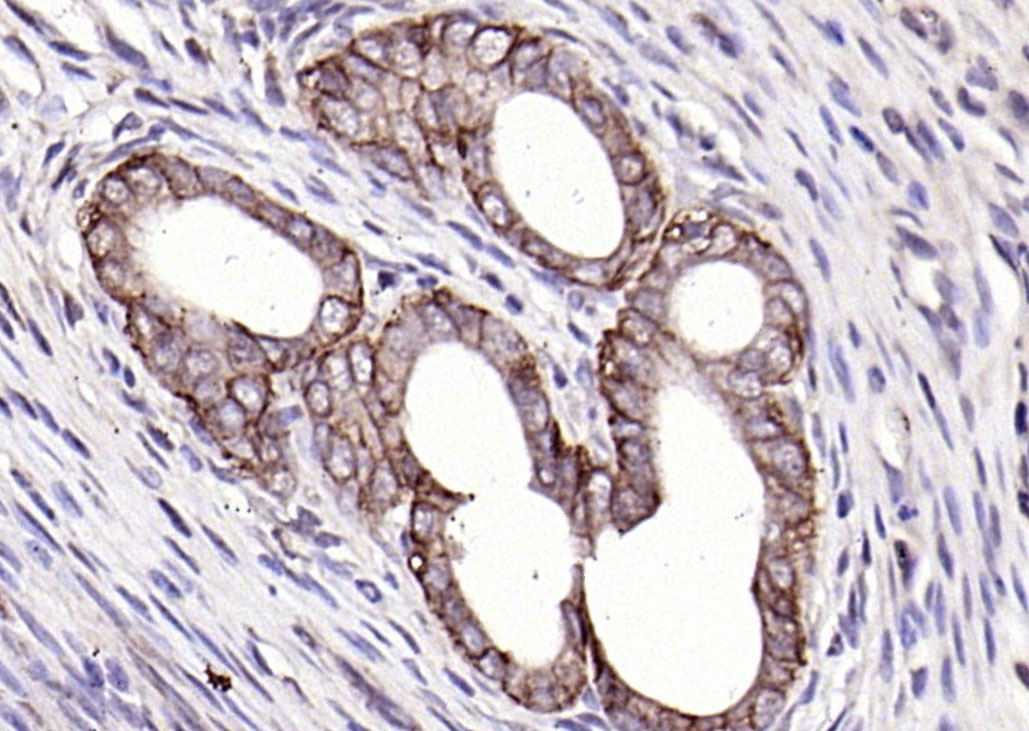 , ,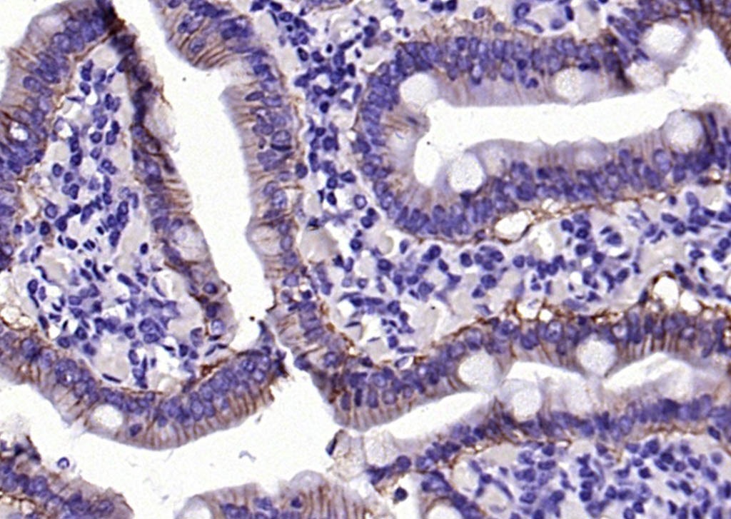 , ,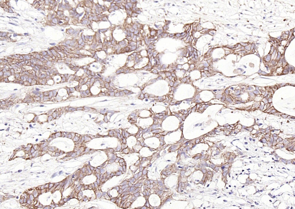 , ,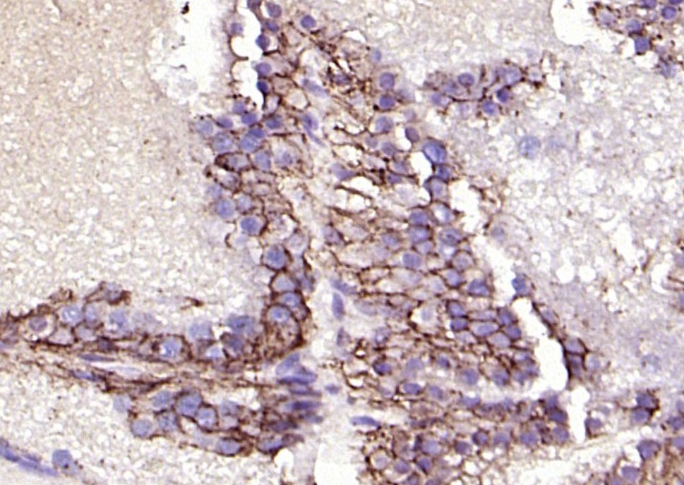 , ,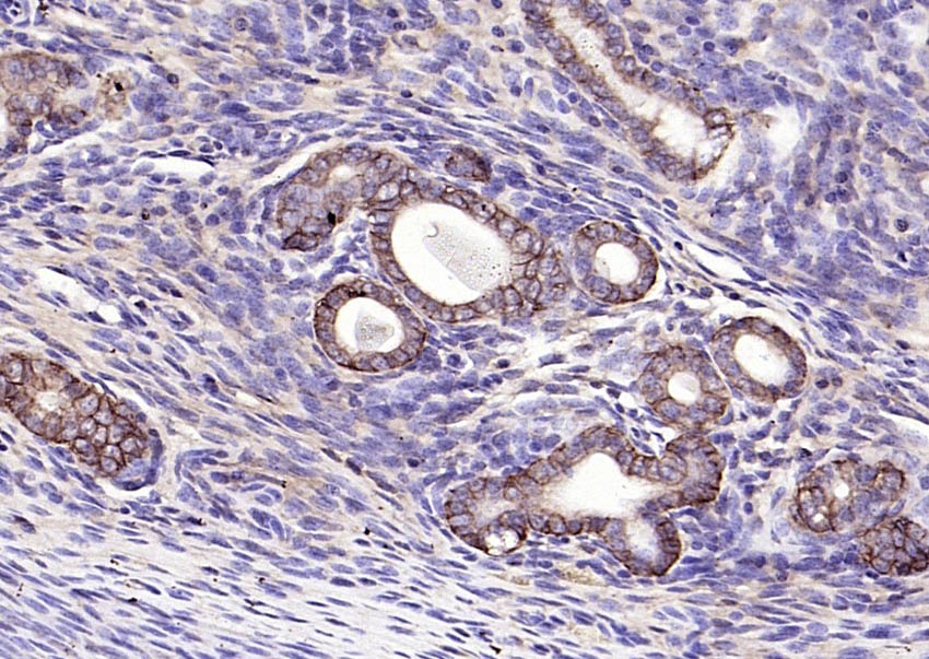 , ,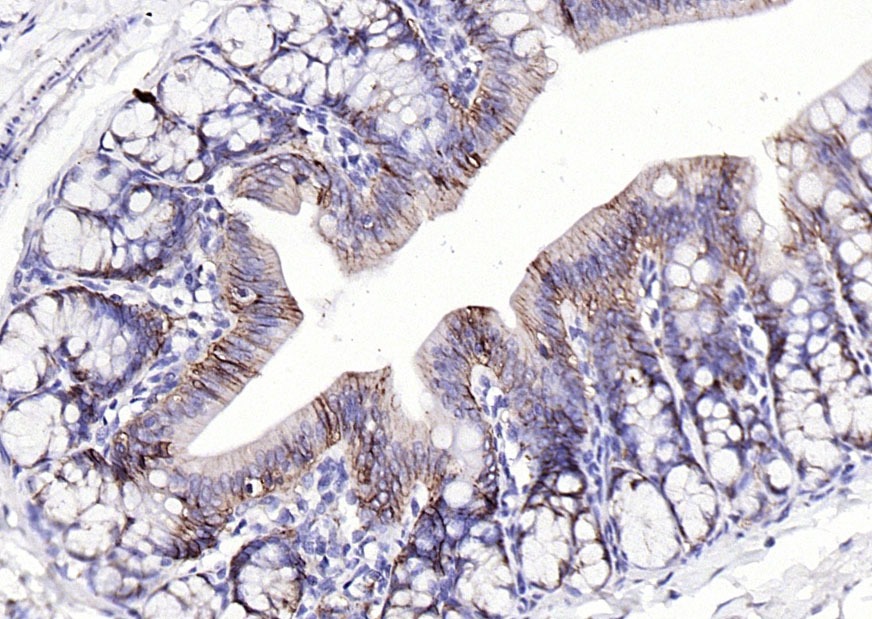 , ,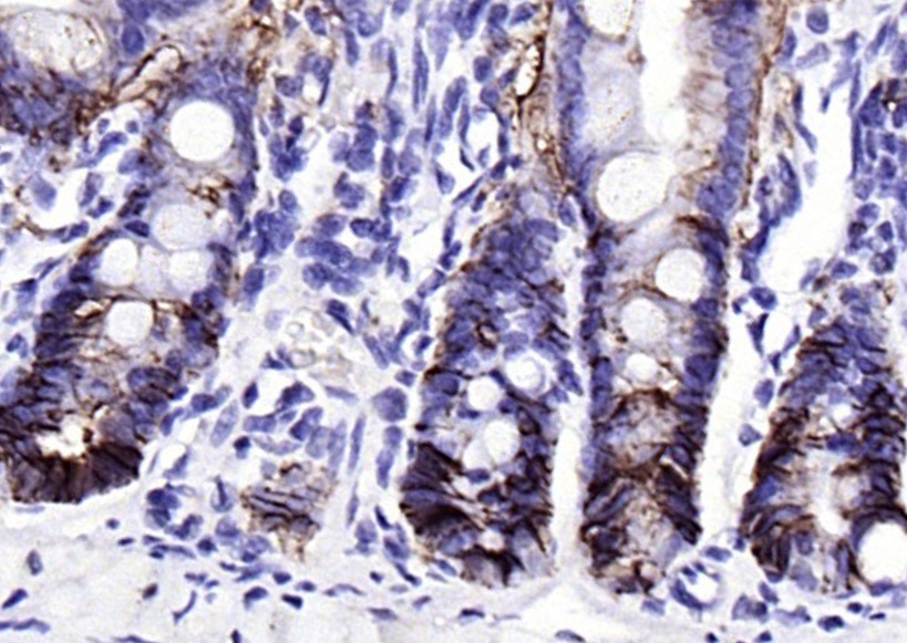 , ,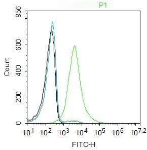 |
| Application | |
| Recommended Dose | WB: 1:500-2000; IHC-P: 1:100-500; IHC-Fr: 1:100-500; IF: 1:100-500; FCM: 1μg/Test |
| Antibody Type | Polyclonal |
| Host Species | Rabbit |
| Subcellular Localization | Lateral cell membrane; Single-pass type I membrane protein. Cell junction, tight junction. Note=Co-localizes with CLDN7 at the lateral cell membrane and tight junction. |
| Tissue Specificity | Highly and selectively expressed by undifferentiated rather than differentiated embryonic stem cells (ESC). Levels rapidly diminish as soon as ESC's differentiate (at protein levels). Expressed in almost all epithelial cell membranes but not on mesodermal |
| Construction | Polyclonal Antibody |
| Purification | Protein A purified |
| Appearance | Liquid |
| Formulation | 0.01M TBS (pH7.4) with 1% BSA, 0.02% Proclin300 and 50% Glycerol. |
| Concentration | 1 mg/mL |
| Research Background | This gene encodes a carcinoma-associated antigen and is a member of a family that includes at least two type I membrane proteins. This antigen is expressed on most normal epithelial cells and gastrointestinal carcinomas and functions as a homotypic calcium-independent cell adhesion molecule. The antigen is being used as a target for immunotherapy treatment of human carcinomas. Mutations in this gene result in congenital tufting enteropathy. [provided by RefSeq, Dec 2008] |
| Immunogen | KLH conjugated synthetic peptide: human EpCAM |
| Antigen Species | Human |
| Gene Name | EPCAM |
| Gene ID | |
| Protein Name | Epithelial cell adhesion molecule |
| Uniprot ID | |
| Biology Area | Epi / Endo-thelial,Tumor Associated |
| Function | May act as a physical homophilic interaction molecule between intestinal epithelial cells (IECs) and intraepithelial lymphocytes (IELs) at the mucosal epithelium for providing immunological barrier as a first line of defense against mucosal infection. Plays a role in embryonic stem cells proliferation and differentiation. Up-regulates the expression of FABP5, MYC and cyclins A and E. |
| Molecular Weight | Theoretical: 35 kDa. Actual: 38 kDa. |
| Stability & Storage | Store at -20°C or -80°C for 12 months. Avoid repeated freeze-thaw cycles. |
| Transport | Shipping with blue ice. |
| Size | Quantity | Unit Price | Amount | Operation |
|---|
 Hello! How can I help you today?
 Copyright © 2015-2026 TargetMol Chemicals Inc. All Rights Reserved.
