
Shopping Cart
Remove All Your shopping cart is currently empty

Synonyms: RP3, microtubule-associated protein, RP/EB family, member 3, EBF3-S, EBF3, EB3
Anti-EB3 Antibody
(1C439)
| Pack Size | Price | USA Stock | Global Stock | Quantity |
|---|---|---|---|---|
| 50 µL | $298 | 7-10 days | 7-10 days | |
| 100 µL | $496 | 7-10 days | 7-10 days |
| Description | Anti-EB3 Antibody (1C439) is a Rabbit antibody targeting EB3. Anti-EB3 Antibody (1C439) can be used in FCM,ICC,IF,IHC,WB. |
| Synonyms | RP3, microtubule-associated protein, RP/EB family, member 3, EBF3-S, EBF3, EB3 |
| Ig Type | IgG |
| Clone | 1C439 |
| Reactivity | Human,Mouse,Rat |
| Verified Activity | 1. Western blot analysis of EB3 on different lysates using anti-EB3 antibody at 1/500 dilution. Positive control: Lane 1: Mouse brain tissue, Lane 2: K562. 2. Immunohistochemical analysis of paraffin-embedded human lung cancer tissue using anti-EB3 antibody. Counter stained with hematoxylin. 3. Immunohistochemical analysis of paraffin-embedded mouse brain tissue using anti-EB3 antibody. Counter stained with hematoxylin. 4. Immunohistochemical analysis of paraffin-embedded mouse fallopian tubes tissue using anti-EB3 antibody. Counter stained with hematoxylin. 5. Immunohistochemical analysis of paraffin-embedded rat cerebellum tissue using anti-EB3 antibody. Counter stained with hematoxylin. 6. ICC staining EB3 in PC-3M cells (green). The nuclear counter stain is DAPI (blue). Cells were fixed in paraformaldehyde, permeabilised with 0.25% Triton X100/PBS. 7. ICC staining EB3 in SH-SY-5Y cells (green). The nuclear counter stain is DAPI (blue). Cells were fixed in paraformaldehyde, permeabilised with 0.25% Triton X100/PBS. 8. ICC staining EB3 in A431 cells (green). The nuclear counter stain is DAPI (blue). Cells were fixed in paraformaldehyde, permeabilised with 0.25% Triton X100/PBS. 9. Flow cytometric analysis of SH-SY-5Y cells with EB3 antibody at 1/100 dilution (red) compared with an unlabelled control (cells without incubation with primary antibody; black). Alexa Fluor 488-conjugated goat anti-rabbit IgG was used as the secondary antibody. 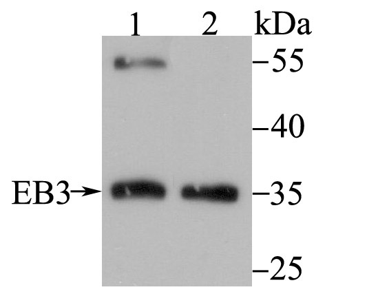 , ,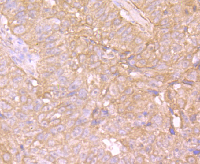 , ,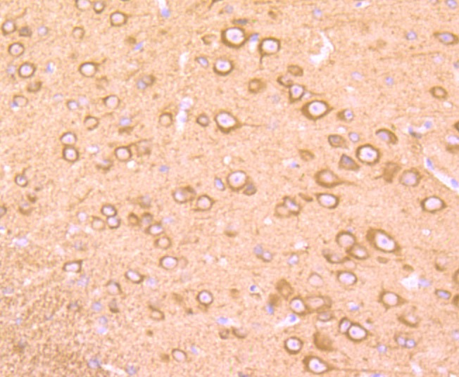 , ,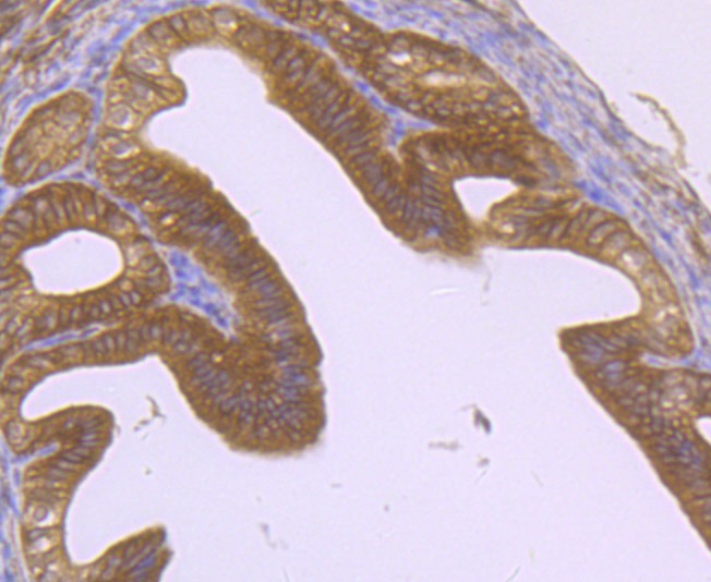 , ,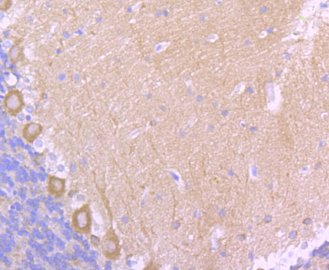 , ,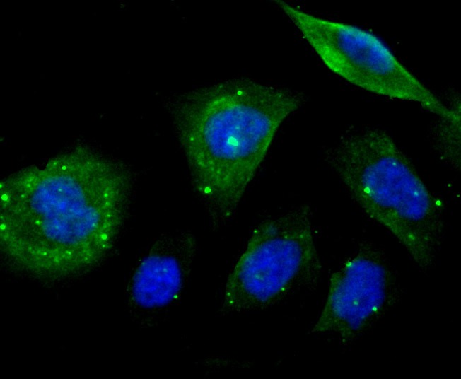 , ,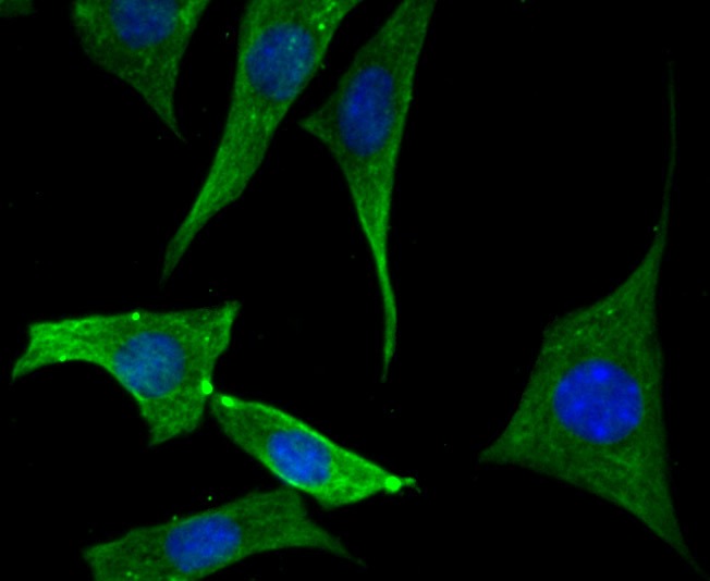 , ,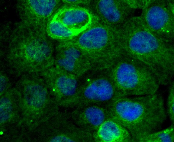 , ,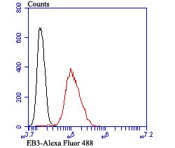 |
| Application | |
| Recommended Dose | WB: 1:500-1000; IHC: 1:50-200; ICC: 1:50-200; FCM: 1:50-100 |
| Antibody Type | Monoclonal |
| Host Species | Rabbit |
| Construction | Recombinant Antibody |
| Purification | ProA affinity purified |
| Appearance | Liquid |
| Formulation | 1*TBS (pH7.4), 1%BSA, 40%Glycerol. Preservative: 0.05% Sodium Azide. |
| Research Background | EB1 (MAPRE2, microtubule-associated protein, RP/EB family, member 2, EB2, RP1) may influence tumorigenesis of colorectal cancers and proliferative control of normal cells. EB1 may belong to the intermediate/early gene family, involved in the signal transduction cascade downstream of the TCR. Colorectal cancer is caused by the pathologic transformation of normal colonic epithelium to an adenomatous polyp, which can become an invasive cancer. APC (adenomatous polyposis coli) is a tumor suppressor gene, the mutation of which is one of the earliest events in colorectal carcinogenesis. A majority of the mutations result in the loss of the carboxy terminus of APC. EB1 has been shown to bind to the carboxy terminal region of APC, which implicates EB1 in APC suppression of colonic cancer. EB1 overexpression may play a role in the development of human esophageal squamous cell carcinoma (ESCC) by affecting APC function and activating the beta-catenin/TCF pathway. EB3 is related to EB1 and likewise associates with the microtubule cytoskeleton. EB3 is expressed predominantly in the central nervous system and preferentially associates with APCL. |
| Conjucates | Unconjugated |
| Immunogen | Recombinant Protein |
| Uniprot ID |
| Molecular Weight | Theoretical: 32 kDa. |
| Stability & Storage | Store at -20°C or -80°C for 12 months. Avoid repeated freeze-thaw cycles. |
| Transport | Shipping with blue ice. |
| Size | Quantity | Unit Price | Amount | Operation |
|---|
 Hello! How can I help you today?
 Copyright © 2015-2026 TargetMol Chemicals Inc. All Rights Reserved.
