
Shopping Cart
Remove All Your shopping cart is currently empty

Synonyms: Cytochrome C, CYCS, CYC
Anti-CYCS Polyclonal Antibody
| Pack Size | Price | USA Stock | Global Stock | Quantity |
|---|---|---|---|---|
| 50 µL | $222 | 7-10 days | 7-10 days | |
| 100 µL | $374 | 7-10 days | 7-10 days | |
| 200 µL | $528 | 7-10 days | 7-10 days |
| Description | Anti-CYCS Polyclonal Antibody is a Rabbit antibody targeting CYCS. Anti-CYCS Polyclonal Antibody can be used in IF, IHC-Fr, IHC-P, WB. |
| Synonyms | Cytochrome C, CYCS, CYC |
| Ig Type | IgG |
| Reactivity | Mouse,Rat,Human |
| Verified Activity | 1. Blank control: HepG2 (blue). Primary Antibody: Rabbit Anti-Cytochrome C antibody (TMAB-00516,Green); Dilution: 1 μg in 100 μL 1X PBS containing 0.5% BSA; Isotype Control Antibody: Rabbit Igg (orange),used under the same conditions; Secondary Antibody: Goat anti-rabbit IgG-FITC (white blue), Dilution: 1:200 in 1 X PBS containing 0.5% BSA. Protocol The cells were fixed with 2% paraformaldehyde for 10 min at 37°C. Primary antibody (TMAB-00516, 1 μg/1x10^6 cells) were incubated for 30 min at room temperature, followed by 1 X PBS containing 0.5% BSA + 10% goat serum (15 min) to block non-specific protein-protein interactions. Then the Goat Anti-rabbit IgG/FITC antibody was added into the blocking buffer mentioned above to react with the primary antibody at 1/200 dilution for 40 min at room temperature. 2. Tissue/cell: SH-SY5Y cell; 4% Paraformaldehyde-fixed; Triton X-100 at room temperature for 20 min; Blocking buffer (normal goat serum) at 37°C for 20 min; Antibody incubation with (Cytochrome C) polyclonal Antibody, Unconjugated (TMAB-00516) 1:100, 90 minutes at 37°C; followed by a FITC conjugated Goat Anti-Rabbit IgG antibody at 37°C for 90 minutes, DAPI (blue) was used to stain the cell nucleus. 3. Sample: Lane 1: Heart (Rat) Lysate at 40 μg Lane 2: Heart (Mouse) Lysate at 40 μg Primary: Anti-Cytochrome C (TMAB-00516) at 1/1000 dilution Secondary: IRDye800CW Goat Anti-Rabbit IgG at 1/20000 dilution Predicted band size: 14.4 kDa Observed band size: 14.4 kDa 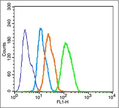 , ,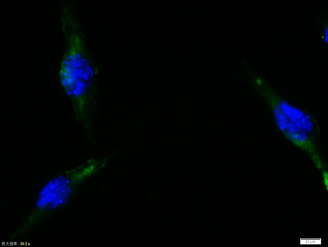 , ,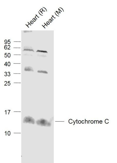 |
| Application | |
| Recommended Dose | IF=1:100-500; IHC-Fr=1:100-500; IHC-P=1:100-500; WB=1:500-2000 |
| Antibody Type | Polyclonal |
| Host Species | Rabbit |
| Subcellular Localization | Mitochondrion intermembrane space. Note=Loosely associated with the inner membrane. |
| Construction | Polyclonal Antibody |
| Purification | Protein A purified |
| Appearance | Liquid |
| Formulation | 0.01M TBS (pH7.4) with 1% BSA, 0.02% Proclin300 and 50% Glycerol. |
| Concentration | 1 mg/mL |
| Research Background | Cytochrome C is an electron transporting protein that resides within the intermembrane space of the mitochondria, where it plays a critical role in the process of oxidative phosphorylation and production of cellular ATP. An increasing amount of interest has been directed toward the role which cytocrome C has been demonstrated to play in apoptotic processes. Following exposure to apoptotic stimuli, cytochrome C is rapidly released from the mitochondria into the cytosol, an event which may be required for the completion of apoptosis in some systems. Cytosolic cytochrome C functions in the activation of caspase 3, an ICE family molecule that is a key effector of apoptosis. |
| Immunogen | KLH conjugated synthetic peptide: human Cytochrome C |
| Antigen Species | Human |
| Gene Name | CYCS |
| Gene ID | |
| Protein Name | Cytochrome c |
| Uniprot ID | |
| Biology Area | Integration of energy metabolism,Cytochrome C,Metabolism,Mitochondrial,Cytochrome C,Cytochromes,Mitochondrial,Energy Metabolism,Integration of energy,Lipases,Apoptosis,Cytochromes,Mitochondrial markers,Oxidative phosphorylation,Energy Metabolism,Mitochondrial |
| Function | Electron carrier protein. The oxidized form of the cytochrome c heme group can accept an electron from the heme group of the cytochrome c1 subunit of cytochrome reductase. Cytochrome c then transfers this electron to the cytochrome oxidase complex, the final protein carrier in the mitochondrial electron-transport chain.Plays a role in apoptosis. Suppression of the anti-apoptotic members or activation of the pro-apoptotic members of the Bcl-2 family leads to altered mitochondrial membrane permeability resulting in release of cytochrome c into the cytosol. Binding of cytochrome c to Apaf-1 triggers the activation of caspase-9, which then accelerates apoptosis by activating other caspases. |
| Molecular Weight | Theoretical: 12 kDa. Actual: 12 kDa. |
| Stability & Storage | Store at -20°C or -80°C for 12 months. Avoid repeated freeze-thaw cycles. |
| Transport | Shipping with blue ice. |
| Size | Quantity | Unit Price | Amount | Operation |
|---|
 Hello! How can I help you today?
 Copyright © 2015-2026 TargetMol Chemicals Inc. All Rights Reserved.
