
Shopping Cart
Remove All Your shopping cart is currently empty

Synonyms: cathepsin B
Anti-Cathepsin B Polyclonal Antibody
| Pack Size | Price | USA Stock | Global Stock | Quantity |
|---|---|---|---|---|
| 50 µL | $220 | 7-10 days | 7-10 days | |
| 100 µL | $374 | 7-10 days | 7-10 days | |
| 200 µL | $528 | 7-10 days | 7-10 days |
| Description | Anti-Cathepsin B Polyclonal Antibody is a Rabbit antibody targeting Cathepsin B. Anti-Cathepsin B Polyclonal Antibody can be used in FCM,IF,IHC-Fr,WB. |
| Synonyms | cathepsin B |
| Ig Type | IgG |
| Reactivity | Human,Mouse,Rat |
| Verified Activity | 1. Sample: Spleen (Mouse) Lysate at 40 μg Primary: Anti-Cathepsin B (TMAB-00297) at 1/300 dilution Secondary: IRDye800CW Goat Anti-Rabbit IgG at 1/20000 dilution Predicted band size: 46/28 kDa Observed band size: 30 kDa 2. Blank control: RSC96 (blue). Primary Antibody: Rabbit Anti-Cathepsin B antibody (TMAB-00297), Dilution: 1 μg in 100 μL 1X PBS containing 0.5% BSA; Isotype Control Antibody: Rabbit Igg (orange),used under the same conditions); Secondary Antibody: Goat anti-rabbit IgG-Pe (white blue), Dilution: 1:200 in 1 X PBS containing 0.5% BSA. Protocol The cells were fixed with 2% paraformaldehyde (10 min). Antibody (TMAB-00297, 5 μg/1x10^6 cells) were incubated for 30 min on the ice, followed by 1 X PBS containing 0.5% BSA + 10% goat serum (15 min) to block non-specific protein-protein interactions. Then the Goat Anti-rabbit IgG/PE antibody was added into the blocking buffer mentioned above to react with the primary antibody of TMAB-00297 at 1/200 dilution for 30 min on ice. 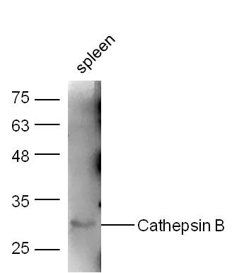 , ,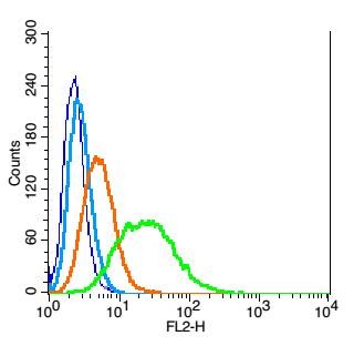 |
| Application | |
| Recommended Dose | WB: 1:500-2000; IHC-Fr: 1:100-500; IF: 1:100-500; FCM: 1µg/Test |
| Antibody Type | Polyclonal |
| Host Species | Rabbit |
| Subcellular Localization | Lysosome. Melanosome. Note=Identified by mass spectrometry in melanosome fractions from stage I to stage IV. |
| Construction | Polyclonal Antibody |
| Purification | Protein A purified |
| Appearance | Liquid |
| Formulation | 0.01M TBS (pH7.4) with 1% BSA, 0.02% Proclin300 and 50% Glycerol. |
| Concentration | 1 mg/mL |
| Research Background | The protein encoded by this gene is a lysosomal cysteine proteinase composed of a dimer of disulfide-linked heavy and light chains, both produced from a single protein precursor. It is also known as amyloid precursor protein secretase and is involved in the proteolytic processing of amyloid precursor protein (APP). Incomplete proteolytic processing of APP has been suggested to be a causative factor in Alzheimer disease, the most common cause of dementia. Overexpression of the encoded protein, which is a member of the peptidase C1 family, has been associated with esophageal adenocarcinoma and other tumors. At least five transcript variants encoding the same protein have been found for this gene. [provided by RefSeq, Jul 2008] |
| Immunogen | KLH conjugated synthetic peptide: human Cathepsin B heavy chain |
| Antigen Species | Human |
| Gene Name | CTSB |
| Gene ID | |
| Protein Name | Cathepsin B |
| Uniprot ID | |
| Biology Area | Cathespins,Cell adhesion,Extracellular matrix,Adhesion molecules ELISA kits,Membrane Proteins,Other Enzymes |
| Function | Thiol protease which is believed to participate in intracellular degradation and turnover of proteins. Has also been implicated in tumor invasion and metastasis. |
| Molecular Weight | Theoretical: 23/28/37 kDa. Actual: 30 kDa. |
| Stability & Storage | Store at -20°C or -80°C for 12 months. Avoid repeated freeze-thaw cycles. |
| Transport | Shipping with blue ice. |
| Size | Quantity | Unit Price | Amount | Operation |
|---|
 Hello! How can I help you today?
 Copyright © 2015-2026 TargetMol Chemicals Inc. All Rights Reserved.
