
Shopping Cart
Remove All Your shopping cart is currently empty

Synonyms:
Anti-BMI-1 Antibody
(8M743)
| Pack Size | Price | USA Stock | Global Stock | Quantity |
|---|---|---|---|---|
| 50 µL | $298 | 7-10 days | 7-10 days | |
| 100 µL | $427 | 7-10 days | 7-10 days |
| Description | Anti-BMI-1 Antibody (8M743) is a Mouse antibody targeting BMI-1. Anti-BMI-1 Antibody (8M743) can be used in FCM,ICC,IHC,WB. |
| Clone | 8M743 |
| Reactivity | Human,Mouse,Rat |
| Verified Activity | 1. Western blot analysis of Bmi1 on different lysates using anti-Bmi1 antibody at 1/1,000 dilution. Positive control: Lane 1: 293T, Lane 2: Jurkat, Lane 3: Hela,, Lane 4: MCF-7, Lane 5: HepG2, Lane 6: NIH/3T3, Lane 7: PC12, Lane 8: Mouse kidney, Lane 9: Human kidney, Lane 10: K562, Lane 11: Human brain. 2. Immunohistochemical analysis of paraffin-embedded human tonsil tissue using anti-Bmi1 antibody. Counter stained with hematoxylin. 3. Immunohistochemical analysis of paraffin-embedded human colon cancer tissue using anti-Bmi1 antibody. Counter stained with hematoxylin. 4. Immunohistochemical analysis of paraffin-embedded human breast cancer tissue using anti-BMI1 antibody. Counter stained with hematoxylin. 5. ICC staining Bmi1 in A549 cells (red). Cells were fixed in paraformaldehyde, permeabilised with 0.25% Triton X100/PBS. 6. ICC staining Bmi1 in Lovo cells (red). Cells were fixed in paraformaldehyde, permeabilised with 0.25% Triton X100/PBS. 7. ICC staining Bmi1 in Hela cells (red). Cells were fixed in paraformaldehyde, permeabilised with 0.25% Triton X100/PBS. 8. Flow cytometric analysis of Hela cells with BMI1 antibody at 1/100 dilution (blue) compared with an unlabelled control (cells without incubation with primary antibody; red). Goat anti mouse IgG (FITC) was used as the secondary antibody. 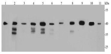 , ,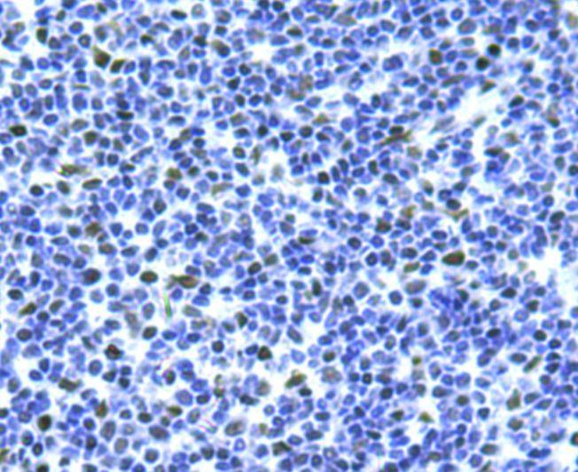 , ,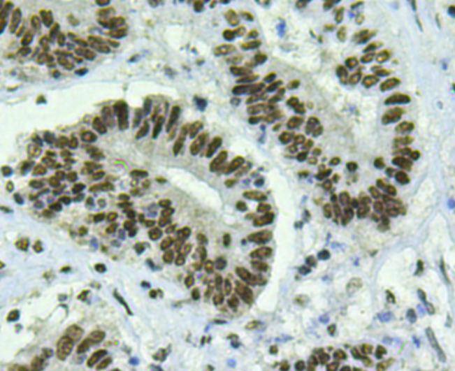 , ,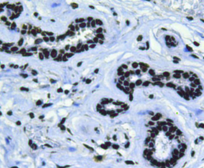 , ,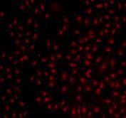 , ,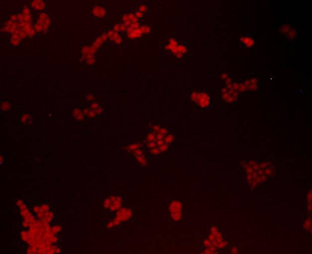 , ,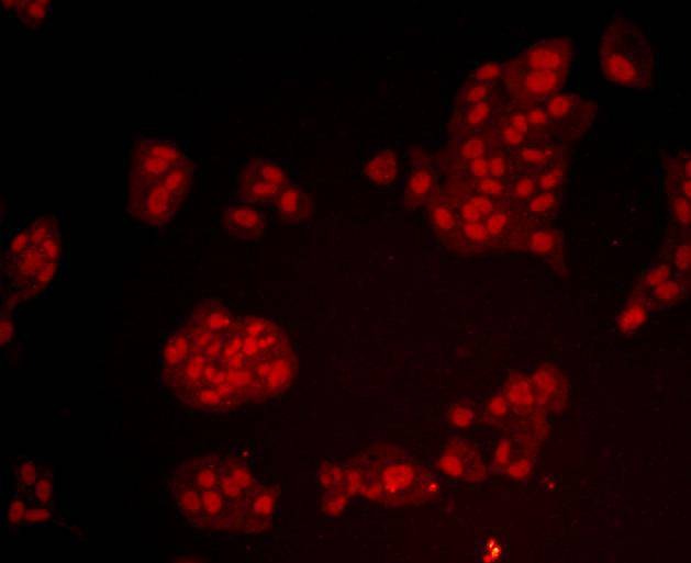 , ,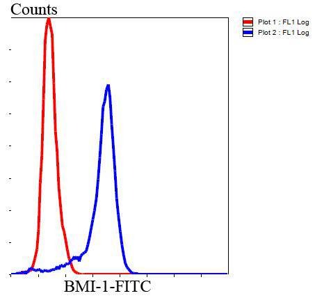 |
| Application | |
| Recommended Dose | WB: 1:1000; IHC: 1:200; ICC: 1:200; FCM: 1:100-200 |
| Antibody Type | Monoclonal |
| Host Species | Mouse |
| Construction | Hybridoma Monoclonal Antibody |
| Purification | ProA affinity purified |
| Appearance | Liquid |
| Formulation | 1*TBS (pH7.4), 1%BSA, 40%Glycerol. Preservative: 0.05% Sodium Azide. |
| Research Background | The Bmi-1 was identified initially as an oncogene that cooperates with c-myc in the generation of B-cell lymphoma. It contributes to the maintenance of cell identity, stem cell self-renewal, cell cycle regulation, and oncogenesis by maintaining the silenced state of genes that promote cell lineage specification, cell death, and cell-cycle arrest. |
| Conjucates | Unconjugated |
| Immunogen | Recombinant Protein: human Bmi1 full sequence |
| Antigen Species | Human |
| Uniprot ID |
| Molecular Weight | Theoretical: 37 kDa. |
| Stability & Storage | Store at -20°C or -80°C for 12 months. Avoid repeated freeze-thaw cycles. |
| Transport | Shipping with blue ice. |
| Size | Quantity | Unit Price | Amount | Operation |
|---|
 Hello! How can I help you today?
 Copyright © 2015-2026 TargetMol Chemicals Inc. All Rights Reserved.
