
Shopping Cart
Remove All Your shopping cart is currently empty

Synonyms:
Anti-ATP5A1 Antibody
(7X811)
| Pack Size | Price | USA Stock | Global Stock | Quantity |
|---|---|---|---|---|
| 50 µL | $296 | 7-10 days | 7-10 days | |
| 100 µL | $498 | 7-10 days | 7-10 days |
| Description | Anti-ATP5A1 Antibody (7X811) is a Rabbit antibody targeting ATP5A1. Anti-ATP5A1 Antibody (7X811) can be used in FCM,ICC/IF,IHC,WB. |
| Ig Type | IgG |
| Clone | 7X811 |
| Reactivity | Human,Mouse,Rat |
| Verified Activity | 1. Immunohistochemical analysis of paraffin-embedded human liver tissue using anti-ATP5A1 antibody. Counter stained with hematoxylin. 2. Immunohistochemical analysis of paraffin-embedded mouse liver tissue using anti-ATP5A1 antibody. Counter stained with hematoxylin. 3. Immunohistochemical analysis of paraffin-embedded mouse heart muscle tissue using anti-ATP5A1 antibody. Counter stained with hematoxylin. 4. ICC staining ATP5A1 in MCF-7 cells (red). The nuclear counter stain is DAPI (blue). Cells were fixed in paraformaldehyde, permeabilised with 0.25% Triton X100/PBS. 5. ICC staining ATP5A1 in HepG2 cells (red). The nuclear counter stain is DAPI (blue). Cells were fixed in paraformaldehyde, permeabilised with 0.25% Triton X100/PBS. 6. ICC staining ATP5A1 in Hela cells (red). The nuclear counter stain is DAPI (blue). Cells were fixed in paraformaldehyde, permeabilised with 0.25% Triton X100/PBS. 7. Flow cytometric analysis of Hela cells with ATP5A1 antibody at 1/50 dilution (red) compared with an unlabelled control (cells without incubation with primary antibody; black). Alexa Fluor 488-conjugated goat anti rabbit IgG was used as the secondary antibody. 8. All lanes use the Antibody at 1:2K dilution for 1 hour at room temperature. 9. Western blot analysis of ATP5A1 expression in HepG2 cell lysate. 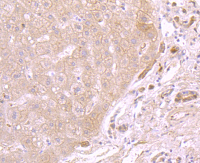 , ,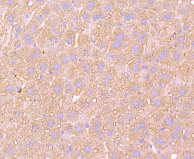 , ,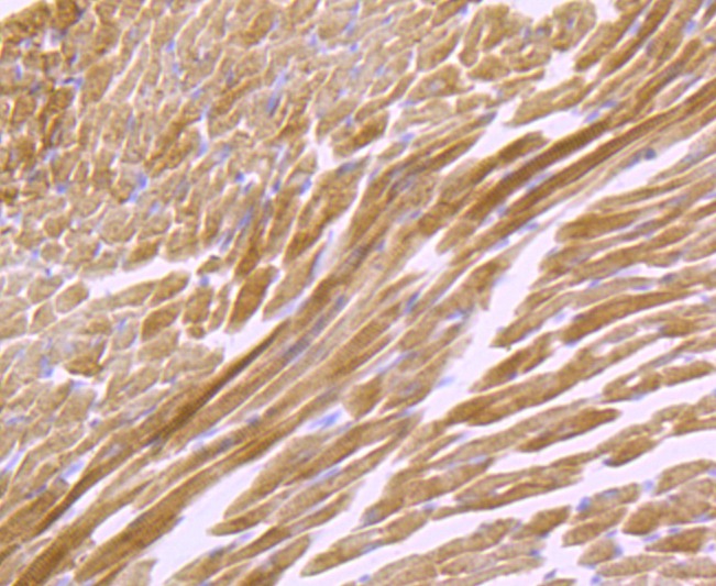 , ,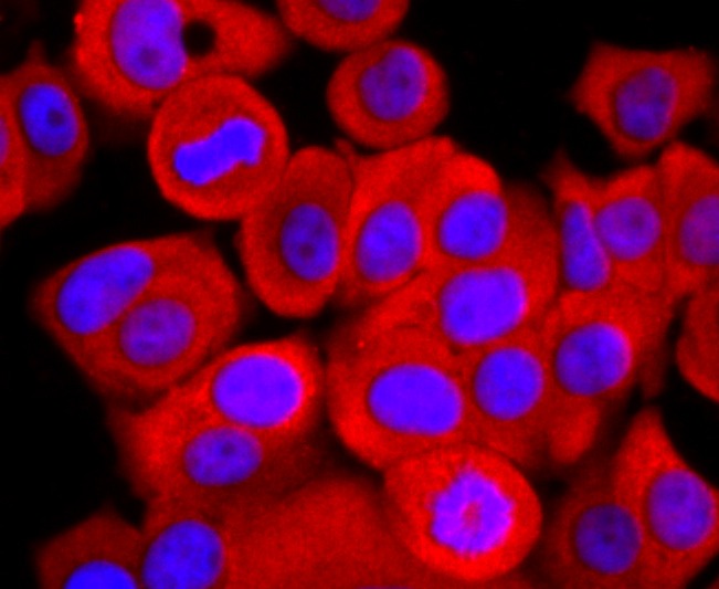 , ,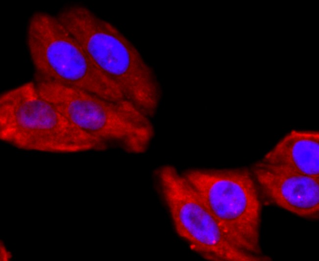 , ,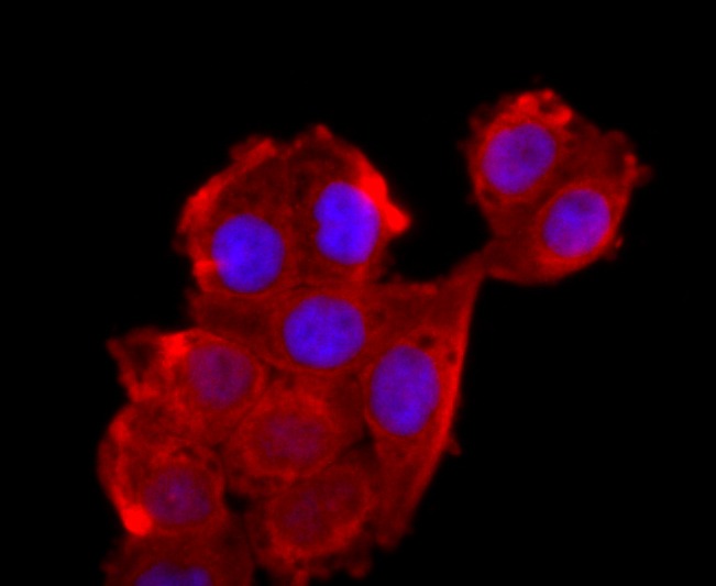 , ,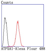 , ,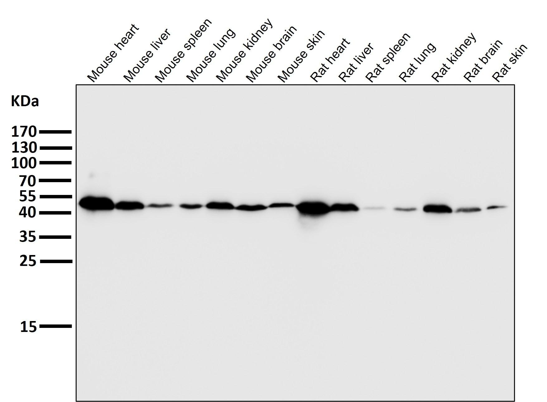 , ,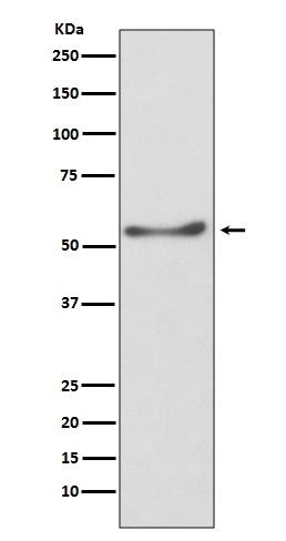 |
| Application | |
| Recommended Dose | WB: 1:1000-2000; IHC: 1:100-200; ICC/IF: 1:50-200; FCM: 1:20-100 |
| Antibody Type | Monoclonal |
| Host Species | Rabbit |
| Construction | Recombinant Antibody |
| Purification | Affinity-chromatography |
| Appearance | Liquid |
| Formulation | Rabbit IgG in 10mM phosphate buffered saline , pH 7.4, 150mM sodium chloride, 0.05% BSA, 0.02% sodium azide and 50% glycerol. |
| Research Background | Mitochondrial ATP synthases (ATPases) transduce the energy contained in membrane electrochemical proton gradients into the energy required for synthesis of high-energy phosphate bonds. ATPases contain two linked complexes: F1, the hydrophilic catalytic core; and F0, the membrane-embedded protein channel. F1 consists of three α chains and three β chains, which are weakly homologous, as well as one γ chain, one δ chain and one e chain. F0 consists of three subunits: a, b and c. The α chain of F1 is a regulatory subunit that contains 509 amino acids. Mitochondrial ATPase α chain (ATP5A) localizes to the mitochondria and catalyzes ATP synthesis. |
| Conjucates | Unconjugated |
| Immunogen | A synthesized peptide: human ATP5A |
| Antigen Species | Human |
| Uniprot ID |
| Molecular Weight | Theoretical: 50 kDa. |
| Stability & Storage | Store at 2°C-8°C for 1 month. Store at -20°C or -80°C for 12 months. Avoid repeated freeze-thaw cycles. |
| Transport | Shipping with blue ice. |
| Size | Quantity | Unit Price | Amount | Operation |
|---|
 Hello! How can I help you today?
 Copyright © 2015-2026 TargetMol Chemicals Inc. All Rights Reserved.
