
Shopping Cart
Remove All Your shopping cart is currently empty

Synonyms: Processed cyclic AMP-dependent transcription factor ATF-6 alpha, FLJ21663, ESTM49, DKFZp686P2194, Cyclic AMP dependent transcription factor ATF 6 alpha, cAMP-dependent transcription factor ATF-6 alpha, ATF6A, ATF6 alpha, ATF6, ATF 6, Activating transcription factor 6 alpha, Activating transcription factor 6
Anti-ATF-6 Polyclonal Antibody
| Pack Size | Price | USA Stock | Global Stock | Quantity |
|---|---|---|---|---|
| 50 µL | $221 | 7-10 days | 7-10 days | |
| 100 µL | $373 | 7-10 days | 7-10 days | |
| 200 µL | $528 | 7-10 days | 7-10 days |
| Description | Anti-ATF-6 Polyclonal Antibody is a Rabbit antibody targeting ATF-6. Anti-ATF-6 Polyclonal Antibody can be used in FCM, ICC/IF, IF, IHC-Fr, IHC-P, WB. |
| Synonyms | Processed cyclic AMP-dependent transcription factor ATF-6 alpha, FLJ21663, ESTM49, DKFZp686P2194, Cyclic AMP dependent transcription factor ATF 6 alpha, cAMP-dependent transcription factor ATF-6 alpha, ATF6A, ATF6 alpha, ATF6, ATF 6, Activating transcription factor 6 alpha, Activating transcription factor 6 |
| Ig Type | IgG |
| Reactivity | Human,Mouse,Rat (predicted:Pig,Cow,Horse,Rabbit) |
| Verified Activity | 1. Tissue/cell: rat brain tissue; 4% Paraformaldehyde-fixed and paraffin-embedded; Antigen retrieval: citrate buffer (0.01M, pH6.0), Boiling bathing for 15 min; Block endogenous peroxidase by 3% Hydrogen peroxide for 30 min; Blocking buffer (normal goat serum) at 37°C for 20 min; Incubation: Anti-ATF6 Polyclonal Antibody, Unconjugated (TMAB-00160) 1:200, overnight at 4°C, followed by conjugation to the secondary antibody and DAb staining. 2. Blank control: A549 (blue). Primary Antibody: Rabbit Anti-ATF6 antibody (TMAB-00160), Dilution: 1 μg in 100 μL 1X PBS containing 0.5% BSA; Isotype Control Antibody: Rabbit Igg (orange),used under the same conditions); Secondary Antibody: Goat anti-rabbit IgG-Pe (white blue), Dilution: 1:200 in 1 X PBS containing 0.5% BSA. Protocol The cells were fixed with 2% paraformaldehyde (10 min). Primary antibody (TMAB-00160, 1 μg/1x10^6 cells) were incubated for 30 min on the ice, followed by 1 X PBS containing 0.5% BSA + 10% goat serum (15 min) to block non-specific protein-protein interactions. Then the Goat Anti-rabbit IgG/PE antibody was added into the blocking buffer mentioned above to react with the primary antibody at 1/200 dilution for 30 min on ice. 3. Sample: Lane 1: Huvec (Human) Cell Lysate at 30 μg Lane 2: Raji (Human) Cell Lysate at 30 μg Lane 3: Jurkat (Human) Cell Lysate at 30 μg Lane 4: A549 (Human) Cell Lysate at 30 μg Lane 5: THP-1 (Human) Cell Lysate at 30 μg Lane 6: 293T (Human) Cell Lysate at 30 μg Lane 7: Du145 (Human) Cell Lysate at 30 μg Primary: Anti-ATF6 (TMAB-00160) at 1/1000 dilution Secondary: IRDye800CW Goat Anti-Rabbit IgG at 1/20000 dilution Predicted band size: 90 kDa Observed band size: 95 kDa 4. Paraformaldehyde-fixed, paraffin embedded (rat brain); Antigen retrieval by boiling in sodium citrate buffer (pH6.0) for 15 min; Block endogenous peroxidase by 3% hydrogen peroxide for 20 min; Blocking buffer (normal goat serum) at 37°C for 30 min; Antibody incubation with (ATF6) Polyclonal Antibody, Unconjugated (TMAB-00160) at 1:200 overnight at 4°C, followed by operating according to SP Kit (Rabbit) instructionsand DAB staining. 5. Paraformaldehyde-fixed, paraffin embedded (mouse brain); Antigen retrieval by boiling in sodium citrate buffer (pH6.0) for 15 min; Block endogenous peroxidase by 3% hydrogen peroxide for 20 min; Blocking buffer (normal goat serum) at 37°C for 30 min; Antibody incubation with (ATF6) Polyclonal Antibody, Unconjugated (TMAB-00160) at 1:200 overnight at 4°C, followed by operating according to SP Kit (Rabbit) instructionsand DAB staining. 6. Blank control: Jurkat. Primary Antibody (green line): Rabbit Anti-ATF6 antibody (TMAB-00160) Dilution: 1 μg/10^6 cells; Isotype Control Antibody (orange line): Rabbit IgG. Secondary Antibody: Goat anti-rabbit IgG-FITC Dilution: 1 μg/test. Protocol The cells were fixed with 4% PFA (10 min at room temperature) and then permeabilized with 90% ice-cold methanol for 20 min at-20°C. The cells were then incubated in 5% BSA to block non-specific protein-protein interactions for 30 min at room temperature. Cells stained with Primary Antibody for 30 min at room temperature. The secondary antibody used for 40 min at room temperature. 7. Blank control: Jurkat. Primary Antibody (green line): Rabbit Anti-ATF6 antibody (TMAB-00160) Dilution: 1 μg/10^6 cells; Isotype Control Antibody (orange line): Rabbit IgG. Secondary Antibody: Goat anti-rabbit IgG-PE Dilution: 1 μg/test. Protocol The cells were fixed with 4% PFA (10 min at room temperature) and then permeabilized with 90% ice-cold methanol for 20 min at-20°C. The cells were then incubated in 5% BSA to block non-specific protein-protein interactions for 30 min at room temperature. Cells stained with Primary Antibody for 30 min at room temperature. The secondary antibody used for 40 min at room temperature. 8. Blank control: Jurkat. Primary Antibody (green line): Rabbit Anti-ATF6 antibody (TMAB-00160) Dilution: 1 μg/10^6 cells; Isotype Control Antibody (orange line): Rabbit IgG. Secondary Antibody: Goat anti-rabbit IgG-FITC Dilution: 1 μg/test. Protocol The cells were fixed with 4% PFA (10 min at room temperature) and then permeabilized with 90% ice-cold methanol for 20 min at-20°C. The cells were then incubated in 5% BSA to block non-specific protein-protein interactions for 30 min at room temperature. Cells stained with Primary Antibody for 30 min at room temperature. The secondary antibody used for 40 min at room temperature. 9. Blank control: Jurkat. Primary Antibody (green line): Rabbit Anti-ATF6 antibody (TMAB-00160) Dilution: 1 μg/10^6 cells; Isotype Control Antibody (orange line): Rabbit IgG. Secondary Antibody: Goat anti-rabbit IgG-FITC Dilution: 1 μg/test. Protocol The cells were fixed with 4% PFA (10 min at room temperature) and then permeabilized with 90% ice-cold methanol for 20 min at-20°C. The cells were then incubated in 5% BSA to block non-specific protein-protein interactions for 30 min at room temperature. Cells stained with Primary Antibody for 30 min at room temperature. The secondary antibody used for 40 min at room temperature. 10. Hela cell; 4% Paraformaldehyde-fixed; Triton X-100 at room temperature for 20 min; Blocking buffer (normal goat serum) at 37°C for 20 min; Antibody incubation with (ATF6) polyclonal Antibody, Unconjugated (TMAB-00160) 1:100, 90 minutes at 37°C; followed by a conjugated Goat Anti-Rabbit IgG antibody at 37°C for 90 minutes, DAPI (blue) was used to stain the cell nucleus. 11. Paraformaldehyde-fixed, paraffin embedded (rat brain); Antigen retrieval by boiling in sodium citrate buffer (pH6.0) for 15 min; Block endogenous peroxidase by 3% hydrogen peroxide for 20 min; Blocking buffer (normal goat serum) at 37°C for 30 min; Antibody incubation with (ATF6) Polyclonal Antibody, Unconjugated (TMAB-00160) at 1:200 overnight at 4°C, followed by operating according to SP Kit (Rabbit) instructionsand DAB staining. 12. Paraformaldehyde-fixed, paraffin embedded (rat brain); Antigen retrieval by boiling in sodium citrate buffer (pH6.0) for 15 min; Block endogenous peroxidase by 3% hydrogen peroxide for 20 min; Blocking buffer (normal goat serum) at 37°C for 30 min; Antibody incubation with (ATF6) Polyclonal Antibody, Unconjugated (TMAB-00160) at 1:200 overnight at 4°C, followed by operating according to SP Kit (Rabbit) instructionsand DAB staining. 13. Paraformaldehyde-fixed, paraffin embedded (rat brain); Antigen retrieval by boiling in sodium citrate buffer (pH6.0) for 15 min; Block endogenous peroxidase by 3% hydrogen peroxide for 20 min; Blocking buffer (normal goat serum) at 37°C for 30 min; Antibody incubation with (ATF6) Polyclonal Antibody, Unconjugated (TMAB-00160) at 1:200 overnight at 4°C, followed by operating according to SP Kit (Rabbit) instructionsand DAB staining. 14. Paraformaldehyde-fixed, paraffin embedded (mouse pancreas); Antigen retrieval by boiling in sodium citrate buffer (pH6.0) for 15 min; Block endogenous peroxidase by 3% hydrogen peroxide for 20 min; Blocking buffer (normal goat serum) at 37°C for 30 min; Antibody incubation with (ATF6) Polyclonal Antibody, Unconjugated (TMAB-00160) at 1:200 overnight at 4°C, followed by operating according to SP Kit (Rabbit) instructionsand DAB staining. 15. Paraformaldehyde-fixed, paraffin embedded (rat pancreas); Antigen retrieval by boiling in sodium citrate buffer (pH6.0) for 15 min; Block endogenous peroxidase by 3% hydrogen peroxide for 20 min; Blocking buffer (normal goat serum) at 37°C for 30 min; Antibody incubation with (ATF6) Polyclonal Antibody, Unconjugated (TMAB-00160) at 1:200 overnight at 4°C, followed by operating according to SP Kit (Rabbit) instructionsand DAB staining. 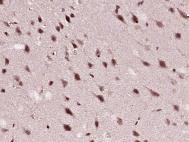 , ,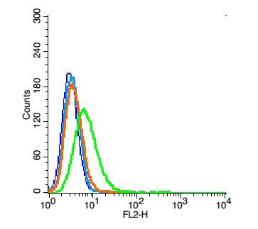 , ,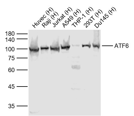 , ,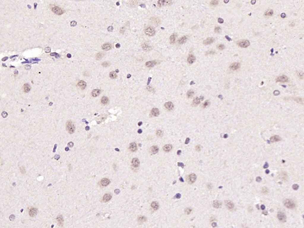 , ,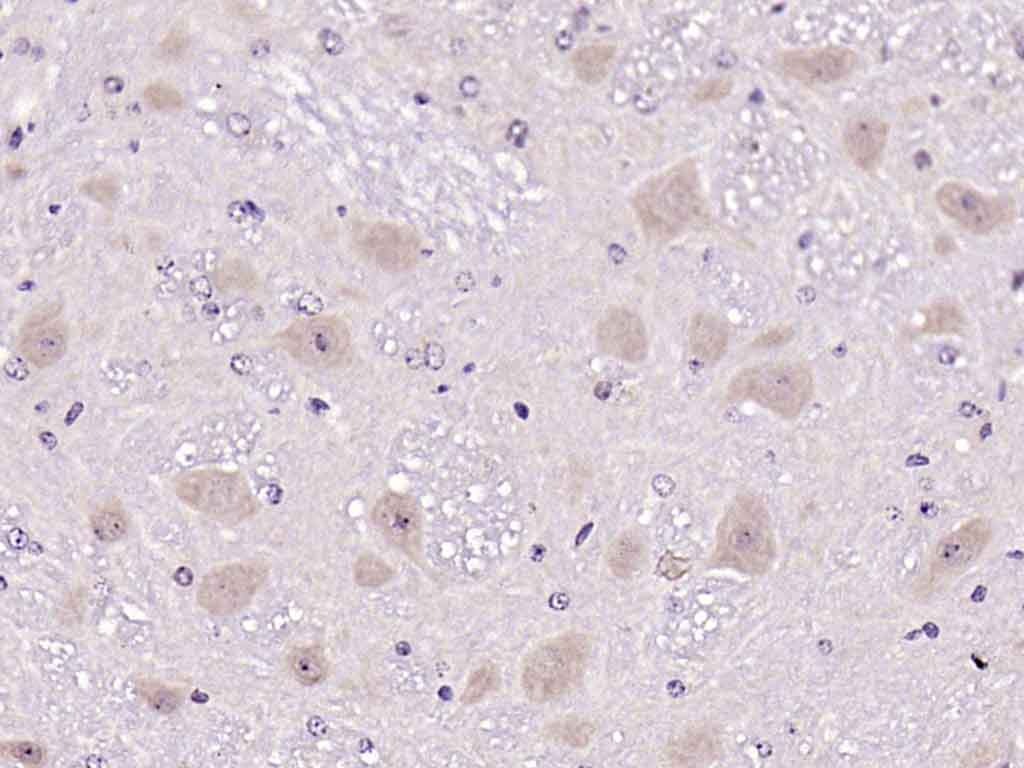 , ,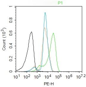 , , , ,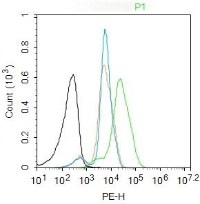 , , , ,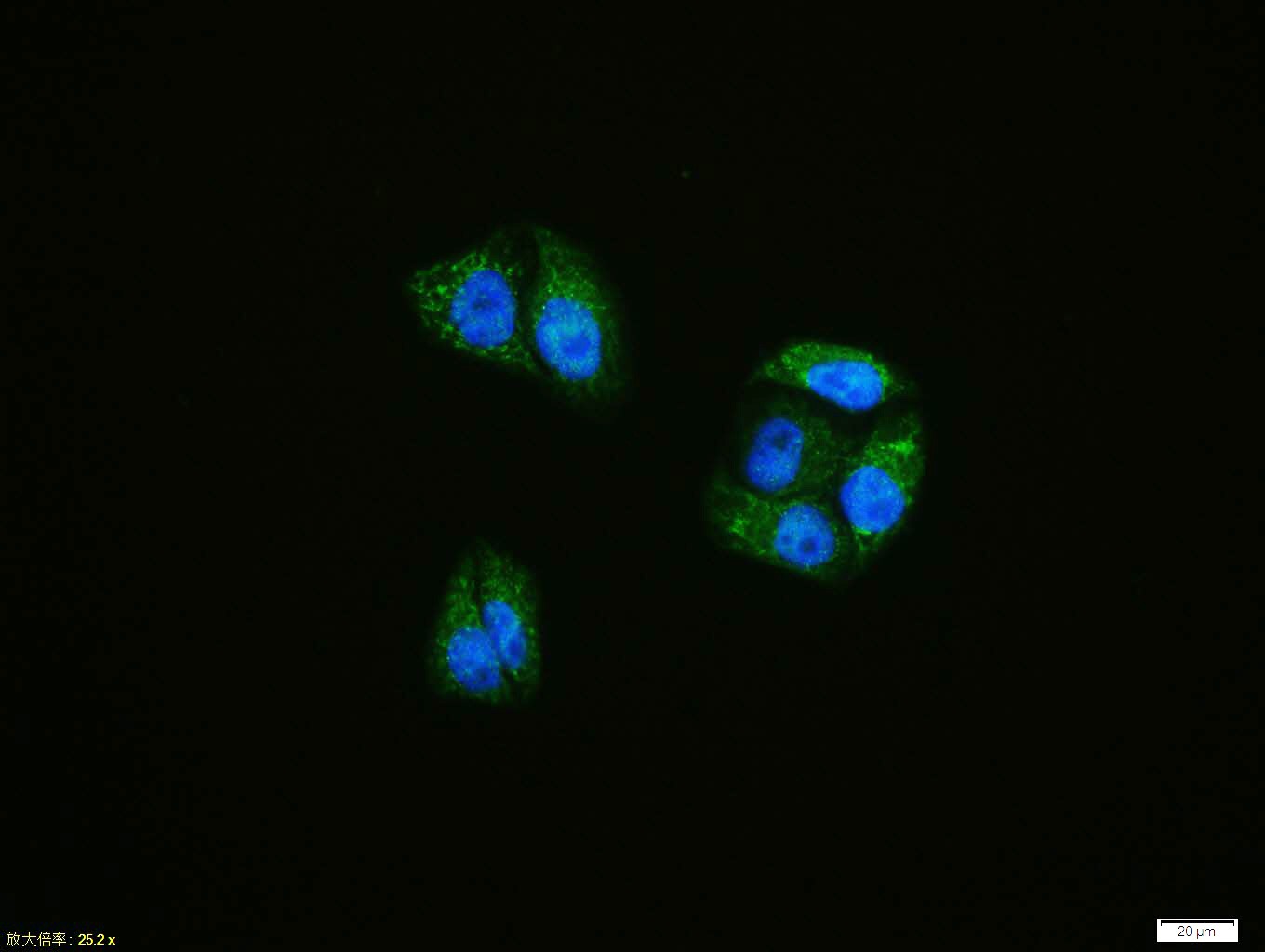 , ,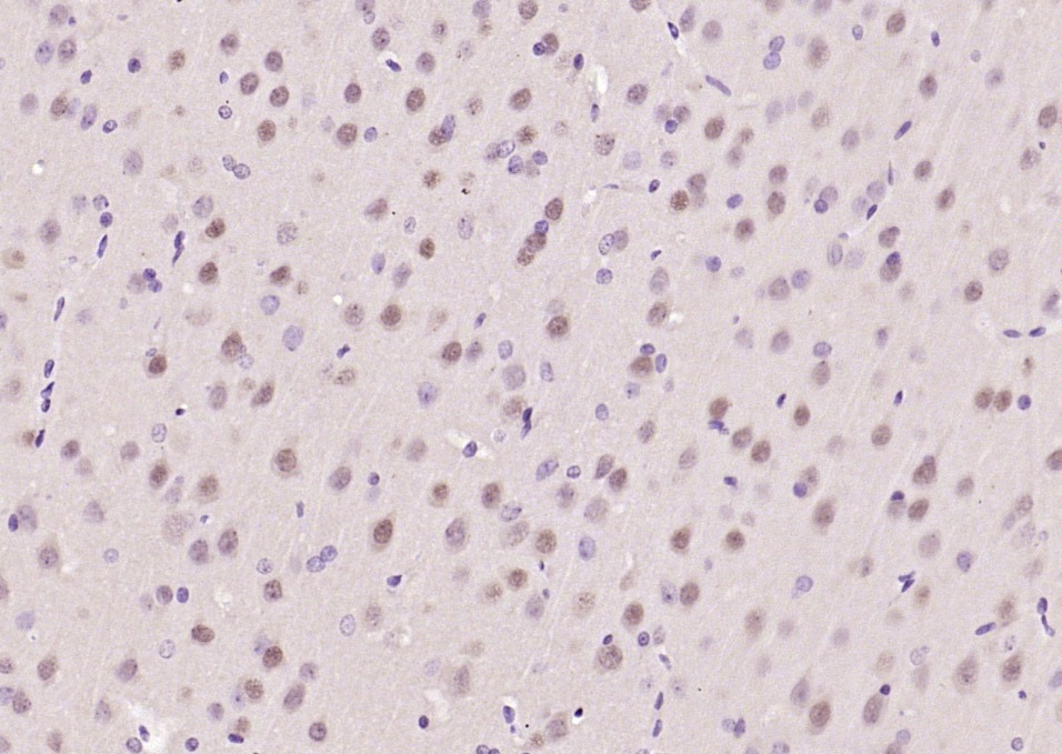 , ,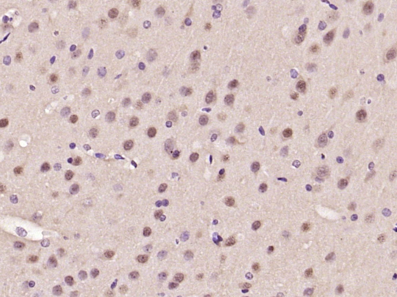 , ,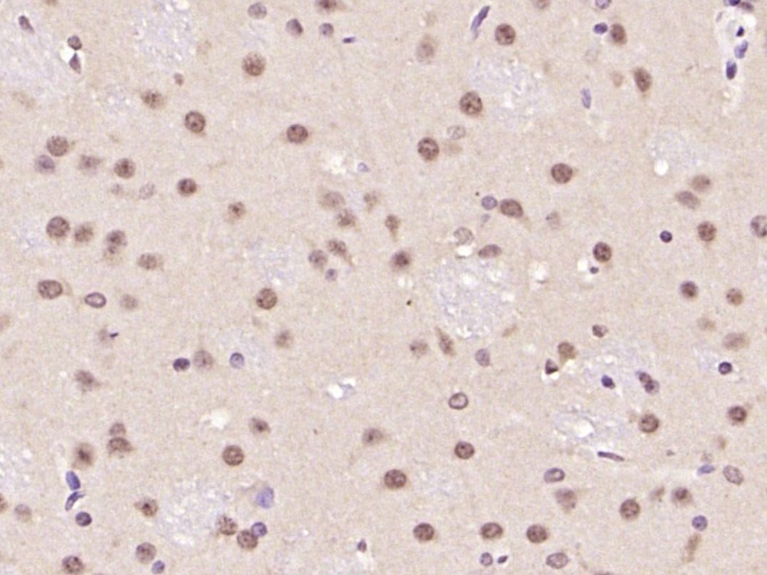 , ,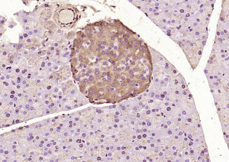 , ,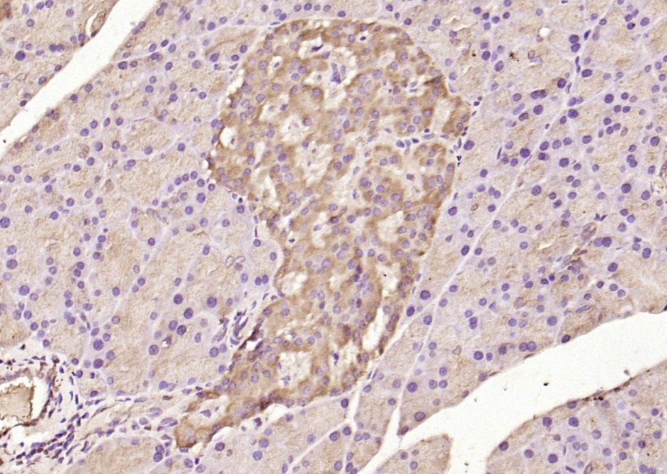 |
| Application | |
| Recommended Dose | FCM=1 μg/Test; ICC/IF=1:100-500; IF=1:100-500; IHC-Fr=1:100-500; IHC-P=1:100-500; WB=1:500-2000 |
| Antibody Type | Polyclonal |
| Host Species | Rabbit |
| Subcellular Localization | Endoplasmic reticulum membrane; Single-pass type II membrane protein. Processed cyclic AMP-dependent transcription factor ATF-6 alpha: Nucleus. Note=Under ER stress the cleaved N-terminal cytoplasmic domain translocates into the nucleus. |
| Tissue Specificity | Ubiquitous. |
| Construction | Polyclonal Antibody |
| Purification | Protein A purified |
| Appearance | Liquid |
| Formulation | 0.01M TBS (pH7.4) with 1% BSA, 0.02% Proclin300 and 50% Glycerol. |
| Concentration | 1 mg/mL |
| Research Background | ATF6 is a transcription factor that acts during endoplasmic reticulum stress by activating unfolded protein response target genes. It binds DNA on the 5'-CCAC[GA]-3'half of the ER stress response element (ERSE) (5'-CCAAT-N(9)-CCAC[GA]-3') and of ERSE II (5'-ATTGG-N-CCACG-3'). Binding to ERSE requires binding of NF-Y to ERSE. ATF6 could also be involved in activation of transcription by the serum response factor. ATF6 exists as a homodimer and heterodimer with ATF6 beta. The dimer interacts with the nuclear transcription factor Y (NF-Y) trimer through direct binding to NF-Y subunit C (NF-YC). It also interacts with the transcription factors GTF2I, YY1 and SRF. Under ER stress the cleaved N-terminal cytoplasmic domain translocates into the nucleus. The basic domain of ATF6 functions as a nuclear localization signal and the basic leucine zipper domain is sufficient for association with the NF-Y trimer and binding to ERSE. During the unfolded protein response an approximately 50 kDa fragment containing the cytoplasmic transcription factor domain is released by proteolysis. The cleavage seems to be performed sequentially by site 1 and site 2 proteases. ATF6 is N glycosylated, phosphorylated in vitro by MAPK14/P38MAPK and belongs to the bZIP family. |
| Immunogen | KLH conjugated synthetic peptide: human ATF6 |
| Antigen Species | Human |
| Gene Name | ATF6 |
| Gene ID | |
| Protein Name | Cyclic AMP-dependent transcription factor ATF-6 alpha |
| Uniprot ID | |
| Biology Area | ChIP antibodies,Leucine Zipper |
| Function | Transcription factor that acts during endoplasmic reticulum stress by activating unfolded protein response target genes. Binds DNA on the 5'-CCAC[GA]-3'half of the ER stress response element (ERSE) (5'-CCAAT-N(9)-CCAC[GA]-3') and of ERSE II (5'-ATTGG-N-CCACG-3'). Binding to ERSE requires binding of NF-Y to ERSE. Could also be involved in activation of transcription by the serum response factor. |
| Molecular Weight | Theoretical: 75 kDa. Actual: 100 kDa. |
| Stability & Storage | Store at -20°C or -80°C for 12 months. Avoid repeated freeze-thaw cycles. |
| Transport | Shipping with blue ice. |
| Size | Quantity | Unit Price | Amount | Operation |
|---|
 Hello! How can I help you today?
 Copyright © 2015-2026 TargetMol Chemicals Inc. All Rights Reserved.
