
Shopping Cart
Remove All Your shopping cart is currently empty

Synonyms: HTX4, ActR-IIB, ACTRIIB, Activin RIIB, activin A receptor, type IIB
Anti-ACVR2B Polyclonal Antibody
| Pack Size | Price | USA Stock | Global Stock | Quantity |
|---|---|---|---|---|
| 50 µL | $222 | 7-10 days | 7-10 days | |
| 100 µL | $372 | 7-10 days | 7-10 days | |
| 200 µL | $529 | 7-10 days | 7-10 days |
| Description | Anti-ACVR2B Polyclonal Antibody is a Rabbit antibody targeting ACVR2B. Anti-ACVR2B Polyclonal Antibody can be used in IF, IHC-Fr, IHC-P, WB. |
| Synonyms | HTX4, ActR-IIB, ACTRIIB, Activin RIIB, activin A receptor, type IIB |
| Ig Type | IgG |
| Reactivity | Human,Mouse (predicted:Rat,Dog,Pig,Cow,Sheep) |
| Verified Activity | 1. Paraformaldehyde-fixed, paraffin embedded (Human kidney); Antigen retrieval by boiling in sodium citrate buffer (pH6.0) for 15 min; Block endogenous peroxidase by 3% hydrogen peroxide for 20 min; Blocking buffer (normal goat serum) at 37°C for 30 min; Antibody incubation with (Activin Receptor Type IIB) Polyclonal Antibody, Unconjugated (TMAB-00052) at 1:200 overnight at 4°C, followed by operating according to SP Kit (Rabbit) instructionsand DAB staining. 2. Sample: K562 (Human) Cell Lysate at 30 μg 293t (Human) Cell Lysate at 30 μg Primary: Anti-Activin Receptor Type IIB (TMAB-00052) at 1/1000 dilution Secondary: IRDye800CW Goat Anti-Rabbit IgG at 1/20000 dilution Predicted band size: 56 kDa Observed band size: 56 kDa 3. Sample: Lane 1: Mouse Heart tissue lysates Lane 2: Mouse Muscle tissue lysates Lane 3: Mouse Testis tissue lysates Lane 4: Mouse Kidney tissue lysates Lane 5: Human K562 cell lysates Lane 6: Human SH-SY5Y cell lysates Lane 7: Human Jurkat cell lysates Lane 8: Human MOLT4 cell lysates Primary: Anti-Activin Receptor Type IIB (TMAB-00052) at 1/1000 dilution Secondary: IRDye800CW Goat Anti-Rabbit IgG at 1/20000 dilution Predicted band size: 56 kDa Observed band size: 60 kDa 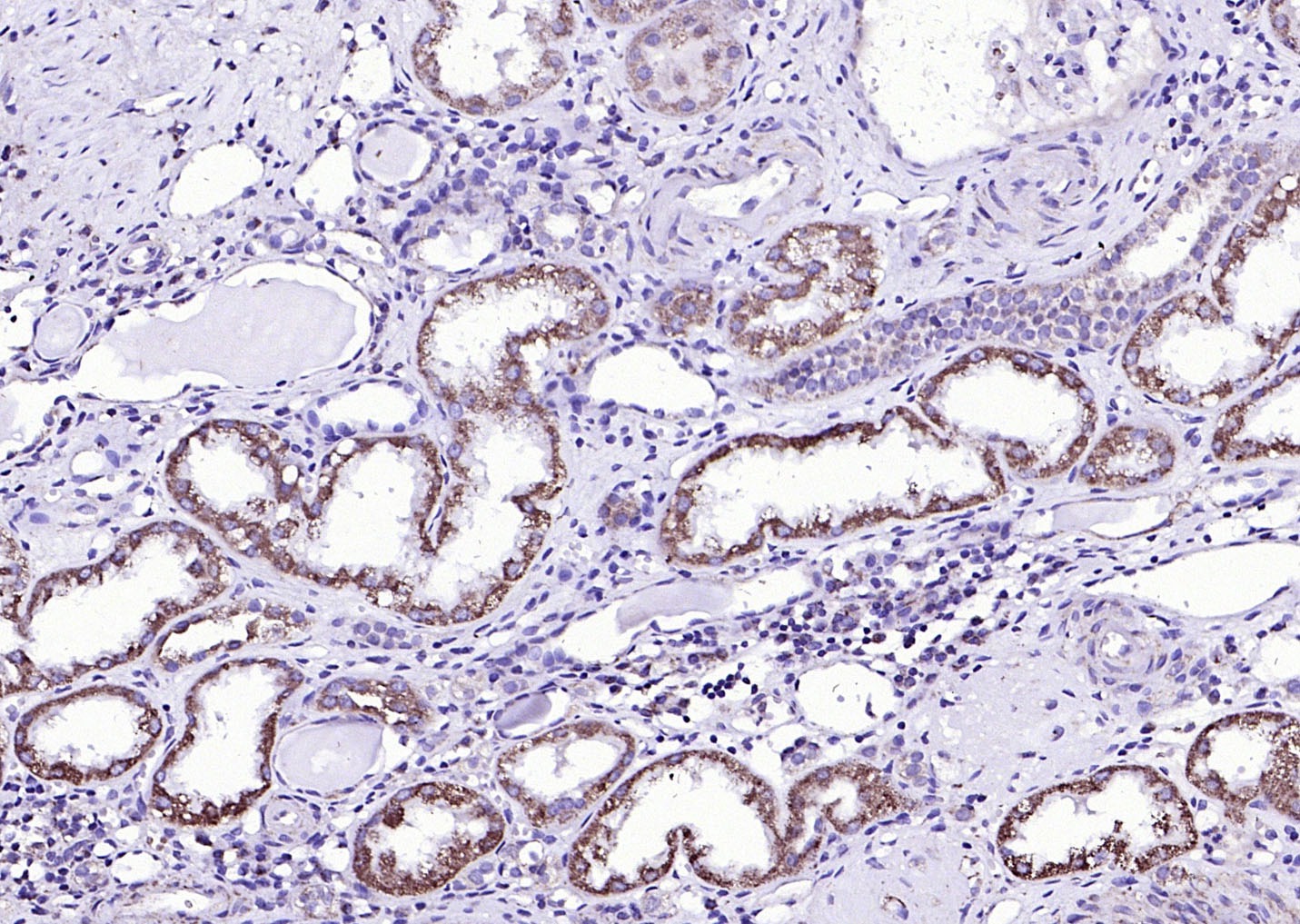 , ,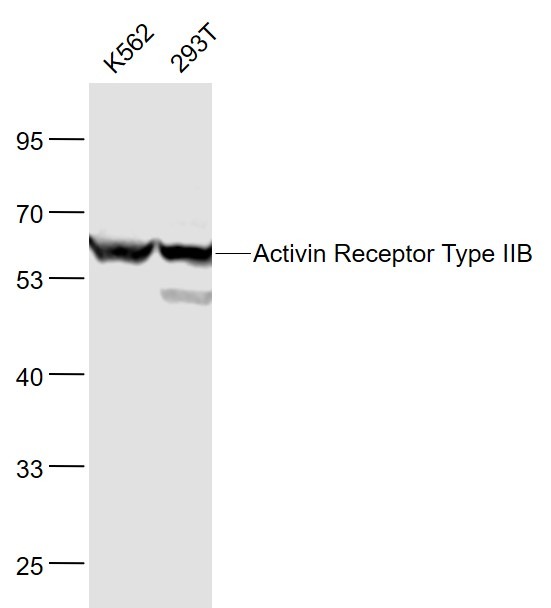 , ,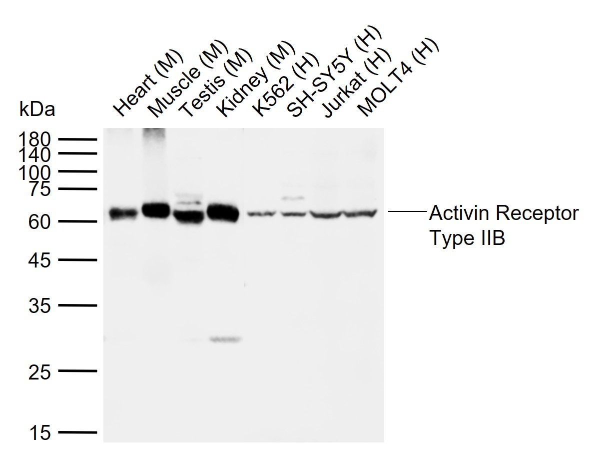 |
| Application | |
| Recommended Dose | WB: 1:500-2000; IHC-P: 1:100-500; IHC-Fr: 1:100-500; IF: 1:100-500 |
| Antibody Type | Polyclonal |
| Host Species | Rabbit |
| Subcellular Localization | Cell membrane; Single-pass type I membrane protein. |
| Construction | Polyclonal Antibody |
| Purification | Protein A purified |
| Appearance | Liquid |
| Formulation | 0.01M TBS (pH7.4) with 1% BSA, 0.02% Proclin300 and 50% Glycerol. |
| Concentration | 1 mg/mL |
| Research Background | Activin receptor type-2B( Activin receptor type IIB; ACTR-IIB ) On ligand binding, forms a receptor complex consisting of two type II and two type I transmembrane serine/threonine kinases. Type II receptors phosphorylate and activate type I receptors which autophosphorylate, then bind and activate SMAD transcriptional regulators. Receptor for activin A, activin B and inhibin A. [Subcellular Location] Membrane; Single-pass type I membrane protein. Belongs to the protein kinase superfamily. TKL Ser/Thr protein kinase family. TGFB receptor subfamily. |
| Immunogen | KLH conjugated synthetic peptide: human ACVR2B/ACTR-IIB |
| Antigen Species | Human |
| Gene Name | ACVR2B |
| Gene ID | |
| Protein Name | Activin receptor type-2B |
| Uniprot ID | |
| Biology Area | Actin Binding Proteins,Other Kinases,Surface Molecules |
| Function | Transmembrane serine/threonine kinase activin type-2 receptor forming an activin receptor complex with activin type-1 serine/threonine kinase receptors (ACVR1, ACVR1B or ACVR1c). Transduces the activin signal from the cell surface to the cytoplasm and is thus regulating many physiological and pathological processes including neuronal differentiation and neuronal survival, hair follicle development and cycling, FSH production by the pituitary gland, wound healing, extracellular matrix production, immunosuppression and carcinogenesis. Activin is also thought to have a paracrine or autocrine role in follicular development in the ovary. Within the receptor complex, the type-2 receptors act as a primary activin receptors (binds activin-A/INHBA, activin-B/INHBB as well as inhibin-A/INHA-INHBA). The type-1 receptors like ACVR1B act as downstream transducers of activin signals. Activin binds to type-2 receptor at the plasma membrane and activates its serine-threonine kinase. The activated receptor type-2 then phosphorylates and activates the type-1 receptor. Once activated, the type-1 receptor binds and phosphorylates the SMAD proteins SMAD2 and SMAD3, on serine residues of the C-terminal tail. Soon after their association with the activin receptor and subsequent phosphorylation, SMAD2 and SMAD3 are released into the cytoplasm where they interact with the common partner SMAD4. This SMAD complex translocates into the nucleus where it mediates activin-induced transcription. Inhibitory SMAD7, which is recruited to ACVR1B through FKBP1A, can prevent the association of SMAD2 and SMAD3 with the activin receptor complex, thereby blocking the activin signal. Activin signal transduction is also antagonized by the binding to the receptor of inhibin-B via the IGSF1 inhibin coreceptor. |
| Molecular Weight | Theoretical: 56 kDa. Actual: 60 kDa. |
| Stability & Storage | Store at -20°C or -80°C for 12 months. Avoid repeated freeze-thaw cycles. |
| Transport | Shipping with blue ice. |
| Size | Quantity | Unit Price | Amount | Operation |
|---|
 Hello! How can I help you today?
 Copyright © 2015-2026 TargetMol Chemicals Inc. All Rights Reserved.
