
Shopping Cart
Remove All Your shopping cart is currently empty

Synonyms: Cell growth-inhibiting gene 46 protein, aortic smooth muscle, Alpha-actin-2, ACTVS, ACTSA, Actin, aortic smooth muscle, ACTA2
Anti-ACTA2 Polyclonal Antibody
| Pack Size | Price | USA Stock | Global Stock | Quantity |
|---|---|---|---|---|
| 50 µL | $222 | 7-10 days | 7-10 days | |
| 100 µL | $372 | 7-10 days | 7-10 days | |
| 200 µL | $527 | 7-10 days | 7-10 days |
| Description | Anti-ACTA2 Polyclonal Antibody is a Rabbit antibody targeting ACTA2. Anti-ACTA2 Polyclonal Antibody can be used in FCM,IF,IHC-Fr,IHC-P,WB. |
| Synonyms | Cell growth-inhibiting gene 46 protein, aortic smooth muscle, Alpha-actin-2, ACTVS, ACTSA, Actin, aortic smooth muscle, ACTA2 |
| Ig Type | IgG |
| Reactivity | Human,Mouse,Rat (predicted:Rabbit) |
| Verified Activity | 1. Blank control (blue line): Hela (blue). Primary Antibody (green line): Rabbit Anti-alpha smooth muscle Actin antibody (TMAB-00047) Dilution: 1 μg/10^6 cells; Isotype Control Antibody (orange line): Rabbit IgG. Secondary Antibody (white blue line): Goat anti-rabbit IgG-FITC Dilution: 1 μg/test. Protocol The cells were fixed with 80% methanol (5 min at-20°C) and then permeabilized with 0.1% PBS-Tween for 20 min at room temperature. Cells stained with Primary Antibody for 30 min at room temperature. The cells were then incubated in 1 X PBS/2% BSA/10% goat serum to block non-specific protein-protein interactions followed by the antibody for 15 min at room temperature. The secondary antibody used for 40 min at room temperature. 2. Blank control: NIH/3T3. Primary Antibody (green line): Rabbit Anti-alpha smooth muscle Actin antibody (TMAB-00047) Dilution: 1 μg/10^6 cells; Isotype Control Antibody (orange line): Rabbit IgG. Secondary Antibody: Goat anti-rabbit IgG-AF488 Dilution: 1 μg/test. Protocol The cells were fixed with 4% PFA (10 min at room temperature) and then permeabilized with 90% ice-cold methanol for 20 min at-20°C. The cells were then incubated in 5% BSA to block non-specific protein-protein interactions for 30 min at room temperature. Cells stained with Primary Antibody for 30 min at room temperature. The secondary antibody used for 40 min at room temperature. 3. Sample: Liver (Rat) Lysate at 40 μg Urinary bladder (Rat) Lysate at 40 μg Stomach (Mouse) Lysate at 40 μg A549 (Cell) Lysate at 30 μg Primary: Anti-alpha smooth muscle Actin (TMAB-00047) at 1/1000 dilution Secondary: IRDye800CW Goat Anti-Rabbit IgG at 1/20000 dilution Predicted band size: 42 kDa Observed band size: 46 kDa 4. Sample: A549 (Human) Cell Lysate at 30 μg NIH/3T3 (Mouse) Cell Lysate at 30 μg Hela (Human) Cell Lysate at 30 μg A431 (Human) Cell Lysate at 30 μg HepG2 (Human) Cell Lysate at 30 μg Stomach (Mouse) Lysate at 40 μg Lung (Mouse) Lysate at 40 μg Skeletal muscle (-) (Mouse) Lysate at 40 μg Primary: Anti-alpha smooth muscle Actin (TMAB-00047) at 1/1000 dilution Secondary: IRDye800CW Goat Anti-Rabbit IgG at 1/20000 dilution Predicted band size: 42 kDa Observed band size: 42 kDa 5. Paraformaldehyde-fixed, paraffin embedded Mouse Stomach; Antigen retrieval by boiling in sodium citrate buffer (pH6.0) for 15 min; Antibody incubation with alpha smooth muscle Actin Polyclonal Antibody, Unconjugated (TMAB-00047) at 1:200 overnight at 4°C, followed by conjugation to the SP Kit (Rabbit) and DAB staining. 6. Paraformaldehyde-fixed, paraffin embedded Human Duodenum; Antigen retrieval by boiling in sodium citrate buffer (pH6.0) for 15 min; Antibody incubation with alpha smooth muscle Actin Polyclonal Antibody, Unconjugated (TMAB-00047) at 1:200 overnight at 4°C, followed by conjugation to the SP Kit (Rabbit) and DAB staining. 7. Paraformaldehyde-fixed, paraffin embedded Human Stomach; Antigen retrieval by boiling in sodium citrate buffer (pH6.0) for 15 min; Antibody incubation with alpha smooth muscle Actin Polyclonal Antibody, Unconjugated (TMAB-00047) at 1:200 overnight at 4°C, followed by conjugation to the SP Kit (Rabbit) and DAB staining. 8. Paraformaldehyde-fixed, paraffin embedded Rat Stomach; Antigen retrieval by boiling in sodium citrate buffer (pH6.0) for 15 min; Antibody incubation with alpha smooth muscle Actin Polyclonal Antibody, Unconjugated (TMAB-00047) at 1:200 overnight at 4°C, followed by conjugation to the SP Kit (Rabbit) and DAB staining. 9. Paraformaldehyde-fixed, paraffin embedded Rat Colon; Antigen retrieval by boiling in sodium citrate buffer (pH6.0) for 15 min; Antibody incubation with alpha smooth muscle Actin Polyclonal Antibody, Unconjugated (TMAB-00047) at 1:200 overnight at 4°C, followed by conjugation to the SP Kit (Rabbit) and DAB staining. 10. Paraformaldehyde-fixed, paraffin embedded Human Cervical Cancer; Antigen retrieval by boiling in sodium citrate buffer (pH6.0) for 15 min; Antibody incubation with alpha smooth muscle Actin Polyclonal Antibody, Unconjugated (TMAB-00047) at 1:200 overnight at 4°C, followed by conjugation to the SP Kit (Rabbit) and DAB staining. 11. Paraformaldehyde-fixed, paraffin embedded Human Colon Cancer; Antigen retrieval by boiling in sodium citrate buffer (pH6.0) for 15 min; Antibody incubation with alpha smooth muscle Actin Polyclonal Antibody, Unconjugated (TMAB-00047) at 1:200 overnight at 4°C, followed by conjugation to the SP Kit (Rabbit) and DAB staining. 12. Blank control: NIH/3T3. Primary Antibody (green line): Rabbit Anti-alpha smooth muscle Actin antibody (TMAB-00047) Dilution: 1 μg/10^6 cells; Isotype Control Antibody (orange line): Rabbit IgG. Secondary Antibody: Goat anti-rabbit IgG-AF488 Dilution: 1 μg/test. Protocol The cells were fixed with 4% PFA (10 min at room temperature) and then permeabilized with 90% ice-cold methanol for 20 min at-20°C. The cells were then incubated in 5% BSA to block non-specific protein-protein interactions for 30 min at room temperature. Cells stained with Primary Antibody for 30 min at room temperature. The secondary antibody used for 40 min at room temperature. 13. Blank control (blue line): Hela (fixed with 70% ethanol (Overnight at 4°C) and then permeabilized with 90% ice-cold methanol for 30 min on ice). Primary Antibody (green line): Rabbit Anti-alpha smooth muscle Actin antibody (TMAB-00047), Dilution: 1 μg/10^6 cells; Isotype Control Antibody (orange line): Rabbit IgG. Secondary Antibody (white blue line): Goat anti-rabbit IgG-FITC, Dilution: 1 μg/test. 14. 25 μg total protein per Lane of various lysates probed with alpha smooth muscle Actin polyclonal antibody, unconjugated (TMAB-00047) at 1:2000 dilution and 4°C overnight incubation. Followed by conjugated secondary antibody incubation at RT for 60 min. 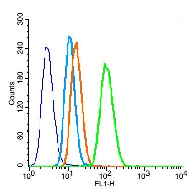 , ,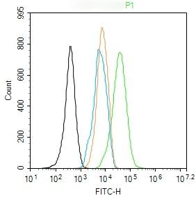 , ,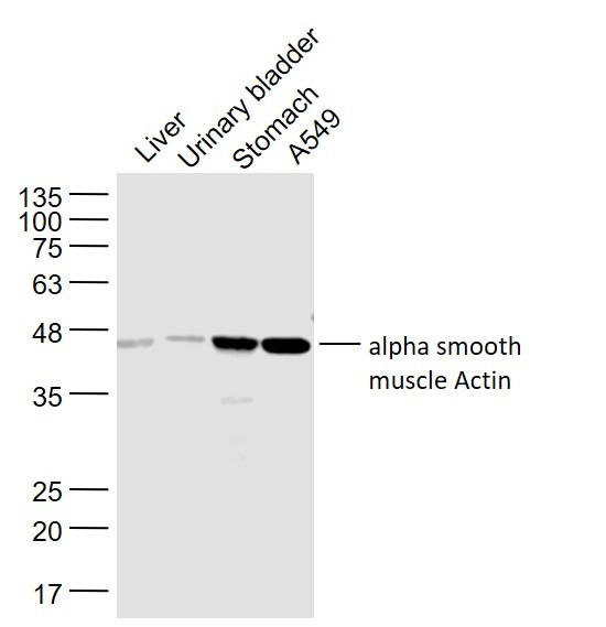 , ,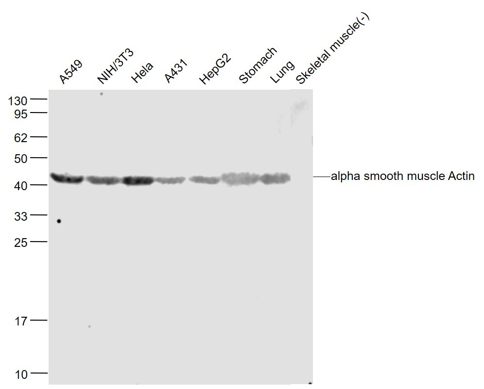 , ,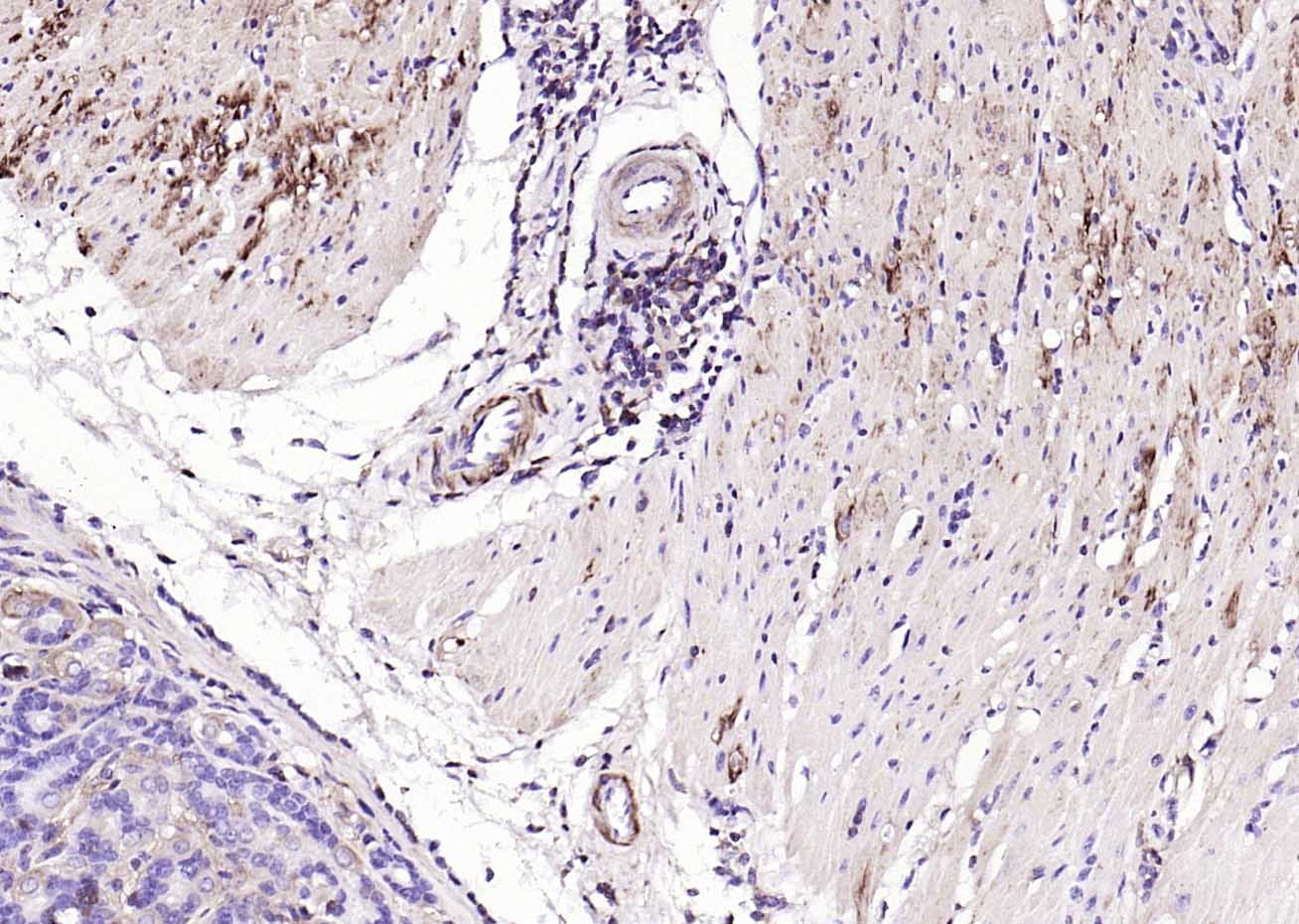 , ,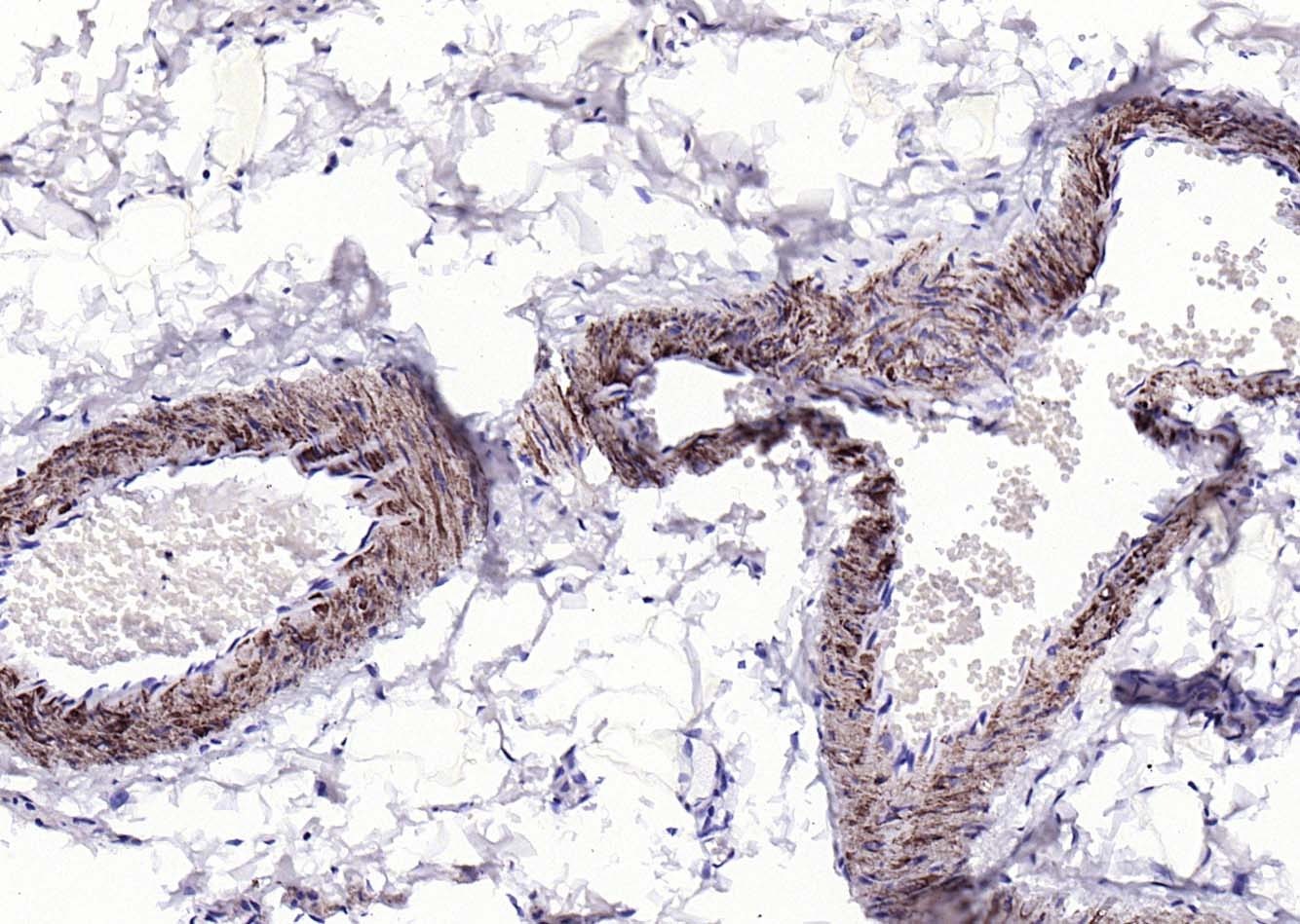 , ,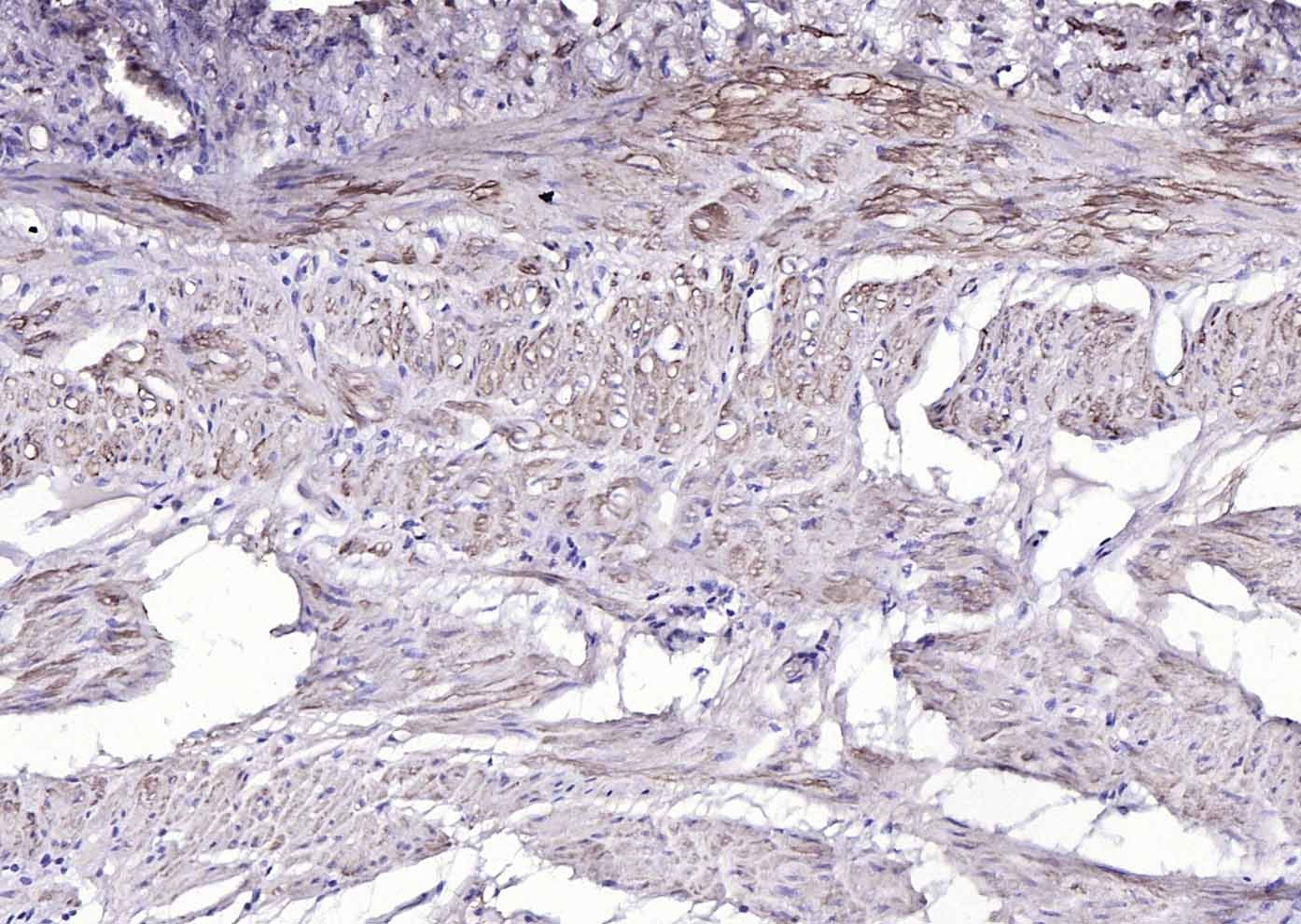 , ,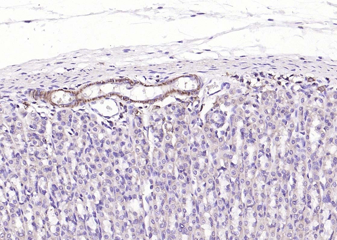 , ,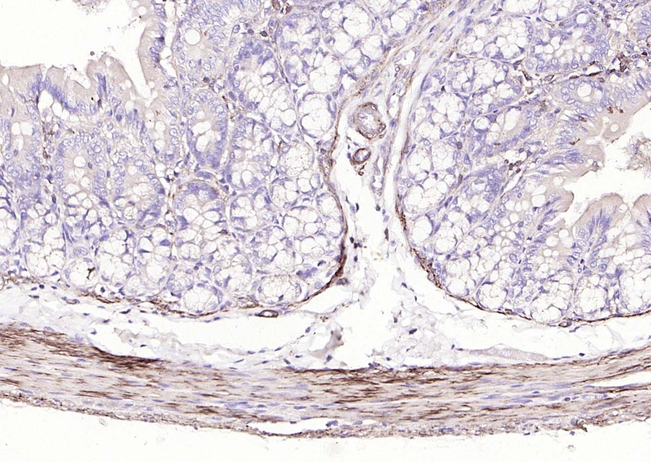 , ,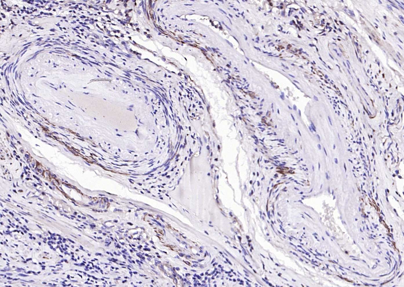 , ,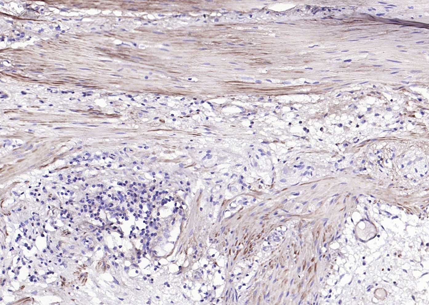 , ,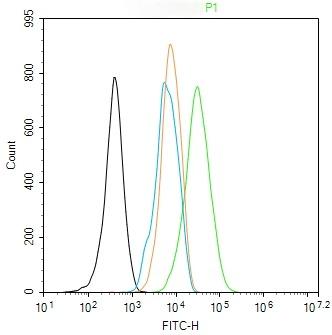 , ,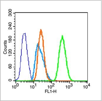 , ,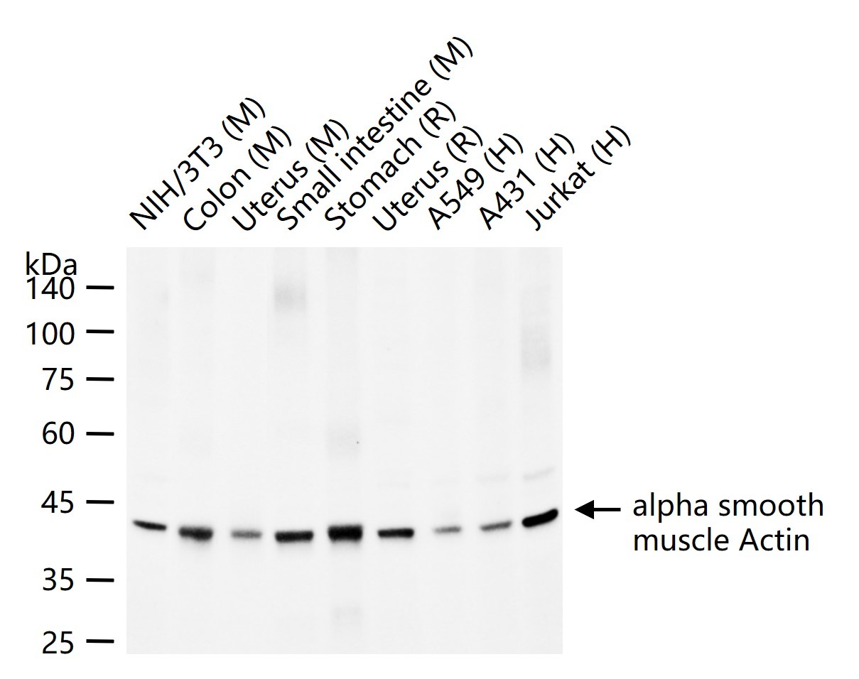 |
| Application | |
| Recommended Dose | WB: 1:1000-5000; IHC-P: 1:100-500; IHC-Fr: 1:100-500; IF: 1:100-500; FCM: 1μg/Test |
| Antibody Type | Polyclonal |
| Host Species | Rabbit |
| Subcellular Localization | Cytoplasm, cytoskeleton. |
| Construction | Polyclonal Antibody |
| Purification | Protein A purified |
| Appearance | Liquid |
| Formulation | 0.01M TBS (pH7.4) with 1% BSA, 0.02% Proclin300 and 50% Glycerol. |
| Concentration | 1 mg/mL |
| Research Background | All eukaryotic cells express Actin, which often constitutes as much as 50% of total cellular protein. Actin filaments can form both stable and labile structures and are crucial components of microvilli and the contractile apparatus of muscle cells. While lower eukaryotes, such as yeast, have only one Actin gene, higher eukaryotes have several isoforms encoded by a family of genes. At least six types of Actin are present in mammalian tissues and fall into three classes. alpha-Actin expression is limited to various types of muscle, whereas beta- and gamma-Actin are the principle constituents of filaments in other tissues. Members of the small GTPase family regulate the organization of the Actin cytoskeleton. Rho controls the assembly of Actin stress fibers and focal adhesion. Rac regulates Actin filament accumulation at the plasma membrane. Cdc42 stimulates formation of filopodia. |
| Immunogen | KLH conjugated synthetic peptide: human Actin alpha |
| Antigen Species | Human |
| Gene Name | ACTA2 |
| Gene ID | |
| Protein Name | Actin, aortic smooth muscle |
| Uniprot ID | |
| Biology Area | Myogenesis,Mesoderm,Actin,alpha smooth muscle Actin,Mesoderm,Arterial,Cytoskeleton,Actins,Smooth Muscle Cells |
| Function | Actins are highly conserved proteins that are involved in various types of cell motility and are ubiquitously expressed in all eukaryotic cells. |
| Molecular Weight | Theoretical: 42 kDa. Actual: 42 kDa. |
| Stability & Storage | Store at -20°C or -80°C for 12 months. Avoid repeated freeze-thaw cycles. |
| Transport | Shipping with blue ice. |
| Size | Quantity | Unit Price | Amount | Operation |
|---|
 Hello! How can I help you today?
 Copyright © 2015-2026 TargetMol Chemicals Inc. All Rights Reserved.
