
Shopping Cart
Remove All Your shopping cart is currently empty
CD4 Protein, Human, Recombinant (hFc) is expressed in HEK293 mammalian cells with hFc tag. The predicted molecular weight is 67.4 kDa and the accession number is A0A4Y5UGE4.
| Pack Size | Price | USA Stock | Global Stock | Quantity |
|---|---|---|---|---|
| 5 μg | $30 | 7-10 days | 7-10 days | |
| 10 μg | $41 | 7-10 days | 7-10 days | |
| 20 μg | $62 | 7-10 days | 7-10 days | |
| 50 μg | $115 | 7-10 days | 7-10 days | |
| 100 μg | $189 | - | In Stock |
| Biological Activity | Measured by the ability of the immobilized protein to support the adhesion of NIH-3T3 mouse embryonic fibroblast cells. When 5×10^4 cells/well are added to CD4-Fc-coated plates (1.25μg/mL, 100 μL/well),approximately 20%-50% cells will adhere after 30 minutes at 37℃. |
| Description | CD4 Protein, Human, Recombinant (hFc) is expressed in HEK293 mammalian cells with hFc tag. The predicted molecular weight is 67.4 kDa and the accession number is A0A4Y5UGE4. |
| Species | Human |
| Expression System | HEK293 Cells |
| Tag | C-hFc |
| Accession Number | P01730 |
| Synonyms | CD4mut,CD4 molecule |
| Construction | A DNA sequence encoding the human CD4 (NP_000607.1) (Met1-Trp390) was expressed with the Fc region of human IgG1 at the C-terminus. Predicted N terminal: Lys 26 |
| Protein Purity | > 95 % as determined by SDS-PAGE. 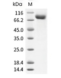 |
| Molecular Weight | 67.4 kDa (predicted) |
| Endotoxin | < 1.0 EU/μg of the protein as determined by the LAL method. |
| Formulation | Lyophilized from a solution filtered through a 0.22 μm filter, containing PBS, pH 7.4. Typically, a mixture containing 5% to 8% trehalose, mannitol, and 0.01% Tween 80 is incorporated as a protective agent before lyophilization. |
| Reconstitution | Reconstituted with sterile deionized water to 0.25 mg/mL. Reconstitution conditions may vary depending on the lot. |
| Stability & Storage | It is recommended to store recombinant proteins at -20°C to -80°C for future use. Lyophilized powders can be stably stored for over 12 months, while liquid products can be stored for 6-12 months at -80°C. For reconstituted protein solutions, the solution can be stored at -20°C to -80°C for at least 3 months. Please avoid multiple freeze-thaw cycles and store products in aliquots. |
| Shipping | In general, lyophilized powders are shipped with blue ice, while solutions are shipped with dry ice. |
| Research Background | T-cell surface glycoprotein CD4, is a single-pass type I membrane protein. CD4 contains three Ig-like C2-type (immunoglobulin-like) domains and one Ig-like V-type (immunoglobulin-like) domain. CD4 is a glycoprotein expressed on the surface of T helper cells, regulatory T cells, monocytes, macrophages, and dendritic cells. The CD4 surface determinant, previously associated as a phenotypic marker for helper/inducer subsets of T lymphocytes, has now been critically identified as the binding/entry protein for human immunodeficiency viruses (HIV). The human CD4 molecule is readily detectable on monocytes, T lymphocytes, and brain tissues. All human tissue sources of CD4 bind radiolabeled gp120 to the same relative degree; however, the murine homologous protein, L3T4, does not bind the HIV envelope protein. CD4 is a co-receptor that assists the T cell receptor (TCR) to activate its T cell following an interaction with an antigen-presenting cell. Using its portion that resides inside the T cell, CD4 amplifies the signal generated by the TCR. CD4 interacts directly with MHC class II molecules on the surface of the antigen-presenting cell via its extracellular domain. The CD4 molecule is currently the object of intense interest and investigation both because of its role in normal T-cell function, and because of its role in HIV infection. CD4 is a primary receptor used by HIV-1 to gain entry into host T cells. HIV infection leads to a progressive reduction of the number of T cells possessing CD4 receptors.Viral protein U (VpU) of HIV-1 plays an important role in downregulation of the main HIV-1 receptor CD4 from the surface of infected cells. Physical binding of VpU to newly synthesized CD4 in the endoplasmic reticulum is an early step in a pathway leading to proteasomal degradation of CD4. Amino acids in both helices found in the cytoplasmic region of VpU in membrane-mimicking detergent micelles experience chemical shift perturbations upon binding to CD4, whereas amino acids between the two helices and at the C-terminus of VpU show no or only small changes, respectively. Paramagnetic spin labels were attached at three sequence positions of a CD4 peptide comprising the transmembrane and cytosolic domains of the receptor. VpU binds to a membrane-proximal region in the cytoplasmic domain of CD4. |
| Size | Quantity | Unit Price | Amount | Operation |
|---|
 Hello! How can I help you today?
 Copyright © 2015-2026 TargetMol Chemicals Inc. All Rights Reserved.