
Shopping Cart
Remove All Your shopping cart is currently empty


Synonyms:
| Pack Size | Price | USA Stock | Global Stock | Quantity |
|---|---|---|---|---|
| 1 mL | $61 | - | In Stock |
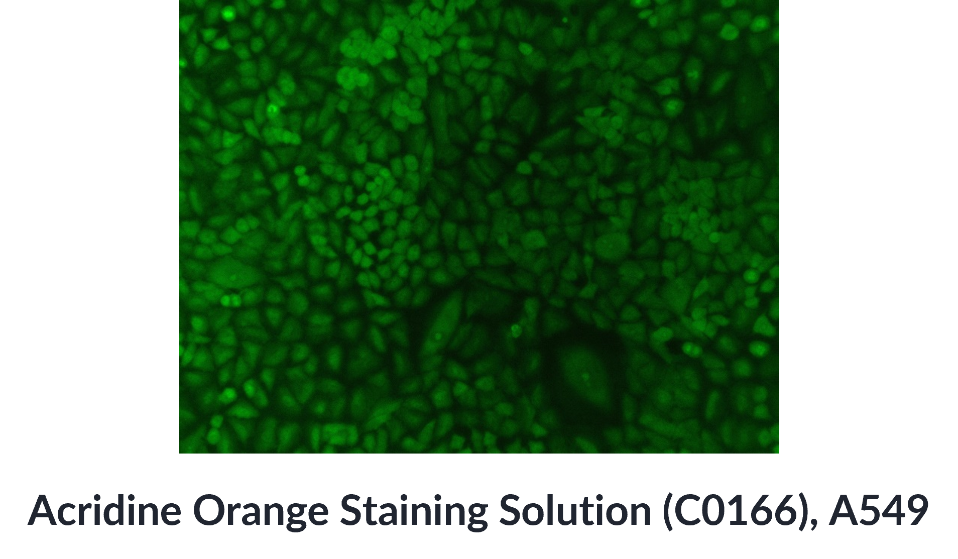
| Acridine Orange Staining Solution | Specifications |
|---|---|
| Ingredient | Acridine Orange |
| CAS | 65-61-2 |
| Conc. | 5 mg/mL |
| Solvent | ddH2O |
1.Exhibits differential staining for DNA and RNA, enabling simultaneous fluorescent labeling of both nucleic acids.
2.Broad application: suitable for nucleic acid labeling in cells, tissues, bacteria, and fungi.
3.Simple operation & high cost-effectiveness.
Apoptosis detection;
Anticancer drug screening;
Cell cycle analysis;
Cell proliferation kinetics analysis.
Dilute the acridine orange stock solution with PBS or ddH2O to prepare a working solution of acridine orange at 0.5-100 μg/mL (perform all steps protected from light). Adjust the exact concentration according to the sample and experimental requirements.
For Adherent Cells
(1)Remove the culture medium from the wells and wash twice with PBS, 2 min each time.
(2)Add acridine orange working solution and stain at room temperature for 5-15 min. Remove the staining solution and wash twice with PBS, 2 min each time.
(3)Add an appropriate amount of PBS to cover the bottom of the wells. Observe under a fluorescence microscope. According to experimental needs, set suitable excitation and emission wavelengths to detect fluorescence.
For green fluorescence: Ex ≈ 488 nm, Em ≈ 530 nm.
For red fluorescence: Ex ≈ 530 nm, Em ≈ 640 nm.
For Suspension Cells
(1)Centrifuge the cell suspension at 600 x g at room temperature for 3 min and discard the supernatant. Resuspend in an appropriate amount of PBS, centrifuge again at 600 x g for 3 min, and discard the supernatant.
(2)Add an appropriate amount of acridine orange working solution and stain at room temperature for 5-15 min.
(3)Either drop directly onto a glass slide, or centrifuge at 600 x g for 3 min, discard the supernatant, resuspend in PBS, and then drop onto a glass slide. Cover with a coverslip and observe directly under a fluorescence microscope, or mount with anti-fade mounting medium before observation. Adjust excitation and emission wavelengths as needed:
Green fluorescence: Ex ≈ 488 nm, Em ≈ 530 nm.
Red fluorescence: Ex ≈ 530 nm, Em ≈ 640 nm.
Store at -20 °C, protected from light for 12 months.
1.When staining with acridine orange, the volume of staining solution is not fixed—just enough to cover the cells.
2.Directly dropping the stained cells onto a glass slide can avoid cell clumping caused by centrifugation; centrifuging to remove the staining solution can help reduce background fluorescence. It is recommended to choose whether to centrifuge after staining based on your experimental needs.
3.Acridine orange solution is light-sensitive, so staining should be performed under dark conditions to minimize fluorescence quenching.
4.The product is for R&D use only, not for diagnostic procedures, food, drug, household or other uses.
5.Please wear a lab coat and disposable gloves.
| Size | Quantity | Unit Price | Amount | Operation |
|---|
 Hello! How can I help you today?
 Copyright © 2015-2026 TargetMol Chemicals Inc. All Rights Reserved.
