
Shopping Cart
Remove All Your shopping cart is currently empty

Synonyms: CTC-85
Anti-XRCC5 Antibody
(9O180)
| Pack Size | Price | USA Stock | Global Stock | Quantity |
|---|---|---|---|---|
| 50 µL | $298 | 7-10 days | 7-10 days | |
| 100 µL | $496 | 7-10 days | 7-10 days |
| Description | Anti-XRCC5 Antibody (9O180) is a Rabbit antibody targeting XRCC5. Anti-XRCC5 Antibody (9O180) can be used in ICC/IF,IHC,IP,WB. |
| Synonyms | CTC-85 |
| Ig Type | IgG |
| Clone | 9O180 |
| Reactivity | Human |
| Verified Activity | 1. Western blot analysis of Ku80 on MCF-7 cells lysates using anti-Ku80 antibody at 1/1,000 dilution. 2. Immunohistochemical analysis of paraffin-embedded human colon cancer tissue using anti-Ku80 antibody. Counter stained with hematoxylin. 3. Immunohistochemical analysis of paraffin-embedded human kideny tissue using anti-Ku80 antibody. Counter stained with hematoxylin. 4. Immunohistochemical analysis of paraffin-embedded human tonsil tissue using anti-Ku80 antibody. Counter stained with hematoxylin. 5. Immunohistochemical analysis of paraffin-embedded human breast carcinoma tissue using anti-Ku80 antibody. Counter stained with hematoxylin. 6. ICC staining Ku80 in Hela cells (green). The nuclear counter stain is DAPI (blue). Cells were fixed in paraformaldehyde, permeabilised with 0.25% Triton X100/PBS. 7. ICC staining Ku80 in A549 cells (green). The nuclear counter stain is DAPI (blue). Cells were fixed in paraformaldehyde, permeabilised with 0.25% Triton X100/PBS. 8. ICC staining Ku80 in SW480 cells (green). The nuclear counter stain is DAPI (blue). Cells were fixed in paraformaldehyde, permeabilised with 0.25% Triton X100/PBS. 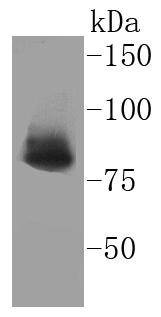 , ,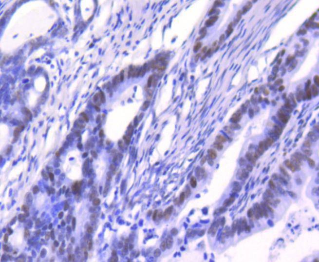 , ,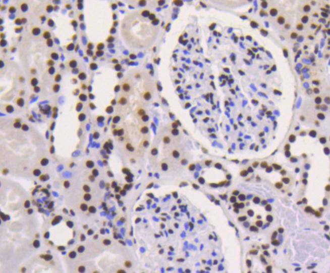 , ,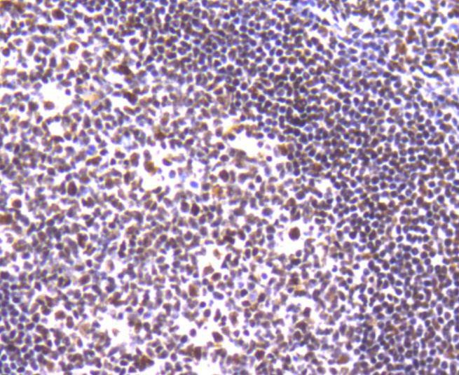 , ,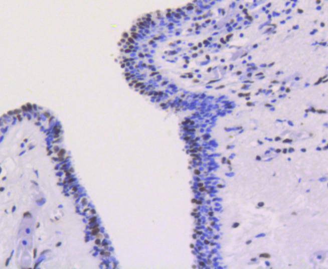 , ,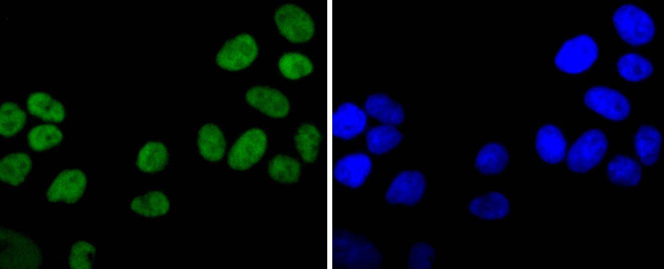 , ,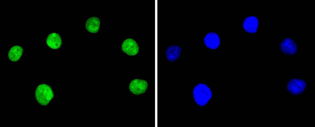 , ,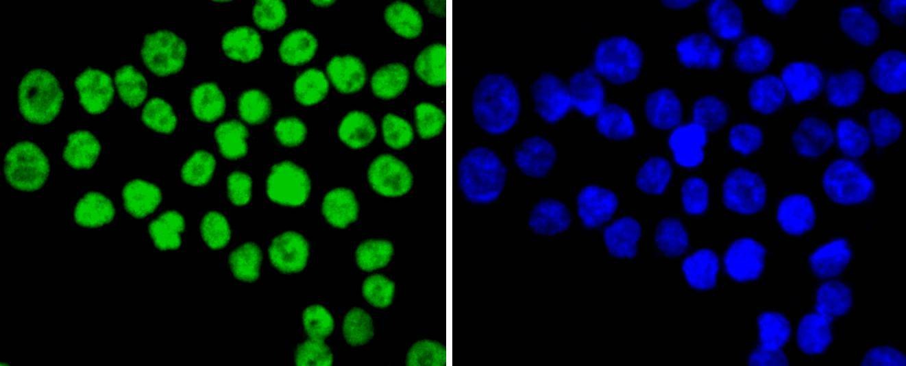 |
| Application | |
| Recommended Dose | WB: 1:1000-2000; IHC: 1:50-200; ICC/IF: 1:50-200 |
| Antibody Type | Monoclonal |
| Host Species | Rabbit |
| Construction | Recombinant Antibody |
| Purification | ProA affinity purified |
| Appearance | Liquid |
| Formulation | 1*TBS (pH7.4), 1%BSA, 40%Glycerol. Preservative: 0.05% Sodium Azide. |
| Research Background | The Ku protein is localized in the nucleus and is composed of subunits referred to as Ku-70 (p70) and Ku-86 (p86) which is also known by the synonym Ku-80 or (p80). Ku was first described as an autoantigen to which antibodies were produced in a patient with scleroderma polymyositis overlap syndrome, and was later found in the sera of patients with other rheumatic diseases. Both subunits of the Ku protein have been cloned, and a number of functions have been proposed for Ku, including cell signaling, DNA replication and transcriptional activation. Ku is involved in Pol II-directed transcription by virtue of its DNA binding activity, serving as the regulatory component of the DNA-associated protein kinase that phosphorylates Pol II and transcription factor Sp. Ku proteins also activate transcription from the U1 small nuclear RNA and the human transferrin receptor gene promoters. A Ku-related protein designated the enhancer 1 binding factor (E1BF), composed of two subunits, has been identified as a positive regulator of RNA polymerase I transcription initiation. |
| Conjucates | Unconjugated |
| Immunogen | Recombinant Protein |
| Uniprot ID |
| Molecular Weight | Theoretical: 83 kDa. |
| Stability & Storage | Store at -20°C or -80°C for 12 months. Avoid repeated freeze-thaw cycles. |
| Transport | Shipping with blue ice. |
| Size | Quantity | Unit Price | Amount | Operation |
|---|
 Hello! How can I help you today?
 Copyright © 2015-2026 TargetMol Chemicals Inc. All Rights Reserved.
