
Shopping Cart
Remove All Your shopping cart is currently empty

Synonyms: Wiskott-Aldrich syndrome protein family member 1, Wiskott Aldrich syndrome protein family member 1, WAVE1, WAVE, WASP family, verprolin homology domain-containing protein 1, WASP family, verprolin homology domain-containing protein, WASP family protein member 1, WASP family member 1, WASP family 1, WASL, WASF1_HUMAN, Wasf1, WAS protein family, member 1, Verprolin homology domain-containing protein 1, Verprolin homology domain containing protein 1, Similar to a plant extensin like protein, SCAR1, scar, Dictyostelium, homology of, 1, Protein WAVE-1v Protein WAVE1, KIAA0269, homology of dictyostelium scar 1, FLJ31482
Anti-WASL Antibody
(8M531)
| Pack Size | Price | USA Stock | Global Stock | Quantity |
|---|---|---|---|---|
| 50 µL | $297 | 7-10 days | 7-10 days | |
| 100 µL | $498 | 7-10 days | 7-10 days |
| Description | Anti-WASL Antibody (8M531) is a Rabbit antibody targeting WASL. Anti-WASL Antibody (8M531) can be used in FCM,ICC,IHC,WB. |
| Synonyms | Wiskott-Aldrich syndrome protein family member 1, Wiskott Aldrich syndrome protein family member 1, WAVE1, WAVE, WASP family, verprolin homology domain-containing protein 1, WASP family, verprolin homology domain-containing protein, WASP family protein member 1, WASP family member 1, WASP family 1, WASL, WASF1_HUMAN, Wasf1, WAS protein family, member 1, Verprolin homology domain-containing protein 1, Verprolin homology domain containing protein 1, Similar to a plant extensin like protein, SCAR1, scar, Dictyostelium, homology of, 1, Protein WAVE-1v Protein WAVE1, KIAA0269, homology of dictyostelium scar 1, FLJ31482 |
| Ig Type | IgG |
| Clone | 8M531 |
| Reactivity | Human,Mouse |
| Verified Activity | 1. Western blot analysis of WASL on mouse Human serum lysates using anti-Ubiquitin antibody at 1/500 dilution. 2. Immunohistochemical analysis of paraffin-embedded human spleen tissue using anti-WASL antibody. Counter stained with hematoxylin. 3. Immunohistochemical analysis of paraffin-embedded human kidney tissue using anti-WASL antibody. Counter stained with hematoxylin. 4. Immunohistochemical analysis of paraffin-embedded mouse colon tissue using anti-WASL antibody. Counter stained with hematoxylin. 5. Immunohistochemical analysis of paraffin-embedded mouse brain tissue using anti-WASL antibody. Counter stained with hematoxylin. 6. Immunohistochemical analysis of paraffin-embedded human colon cancer tissue using anti-WASL antibody. Counter stained with hematoxylin. 7. ICC staining WASL in SK-Br-3 cells (green). The nuclear counter stain is DAPI (blue). Cells were fixed in paraformaldehyde, permeabilised with 0.25% Triton X100/PBS. 8. ICC staining WASL in SH-SY5Y cells (green). The nuclear counter stain is DAPI (blue). Cells were fixed in paraformaldehyde, permeabilised with 0.25% Triton X100/PBS. 9. Flow cytometric analysis of K562 cells with WASL antibody at 1/100 dilution (red) compared with an unlabelled control (cells without incubation with primary antibody; black). 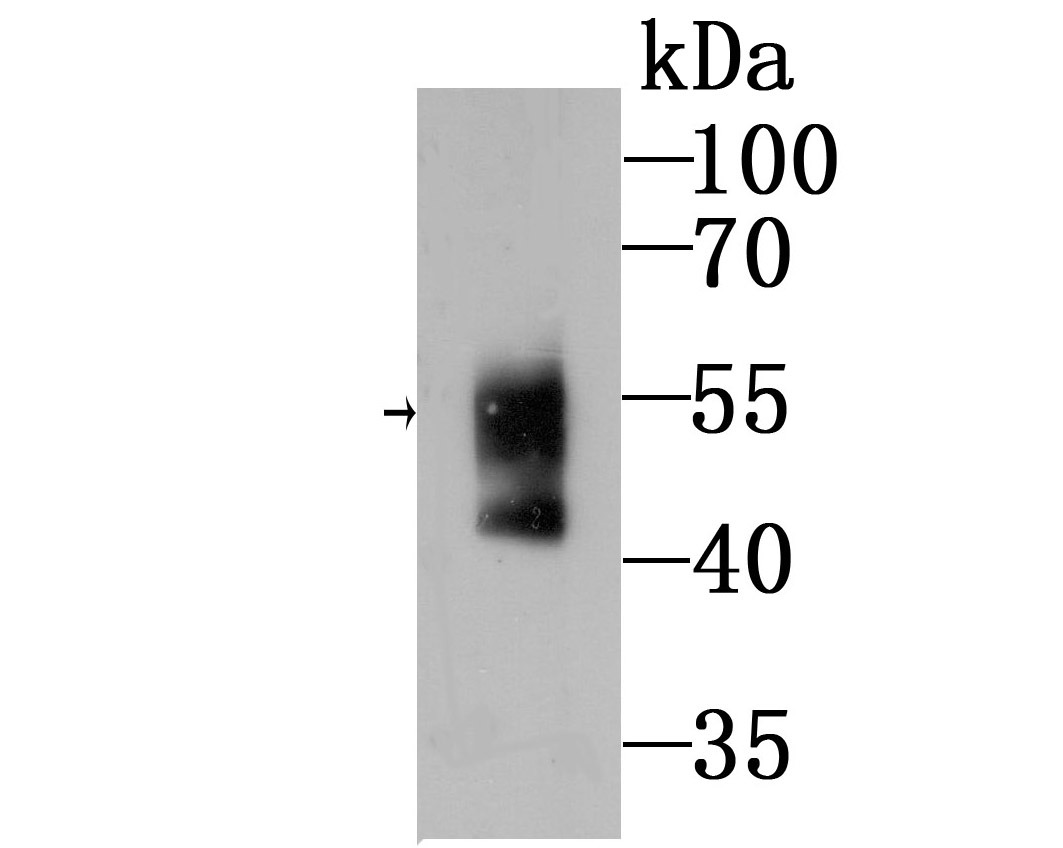 , ,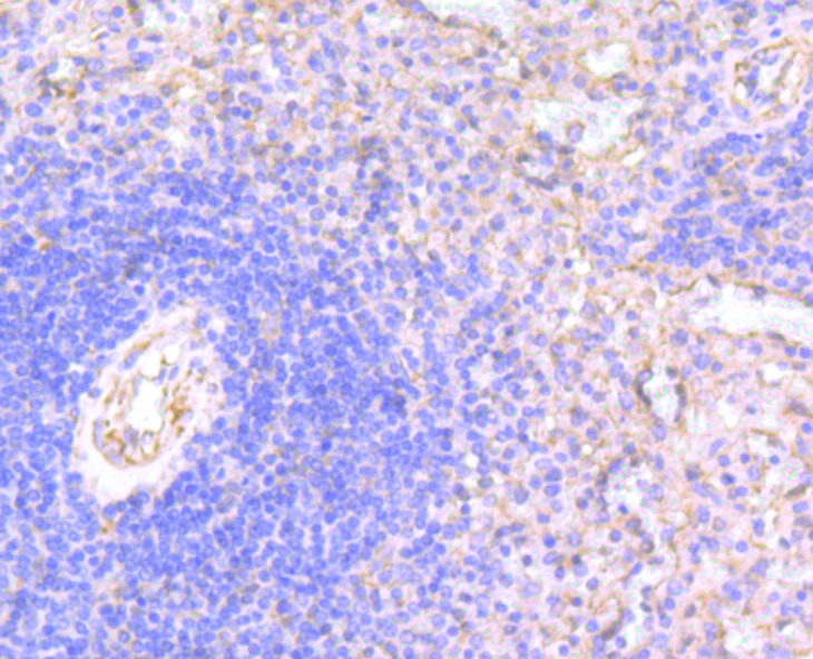 , ,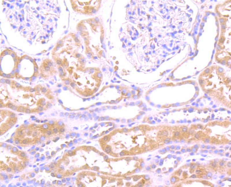 , ,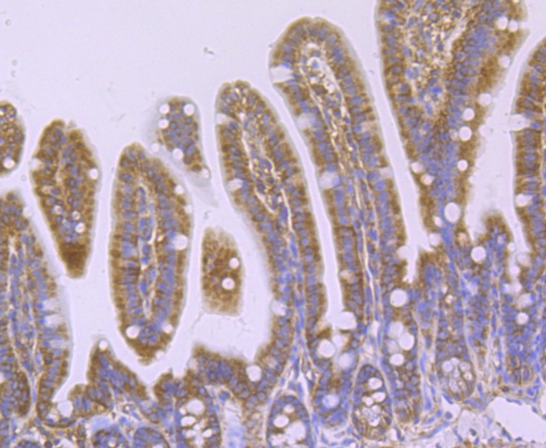 , ,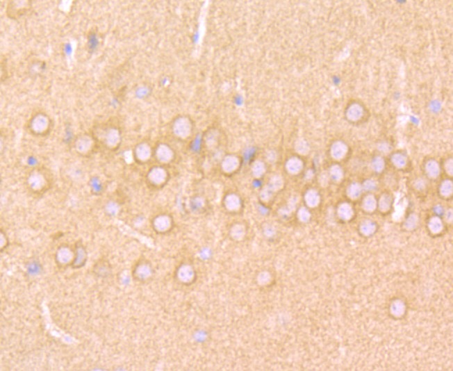 , ,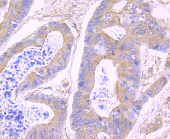 , ,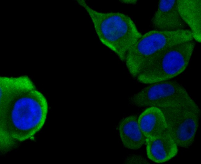 , ,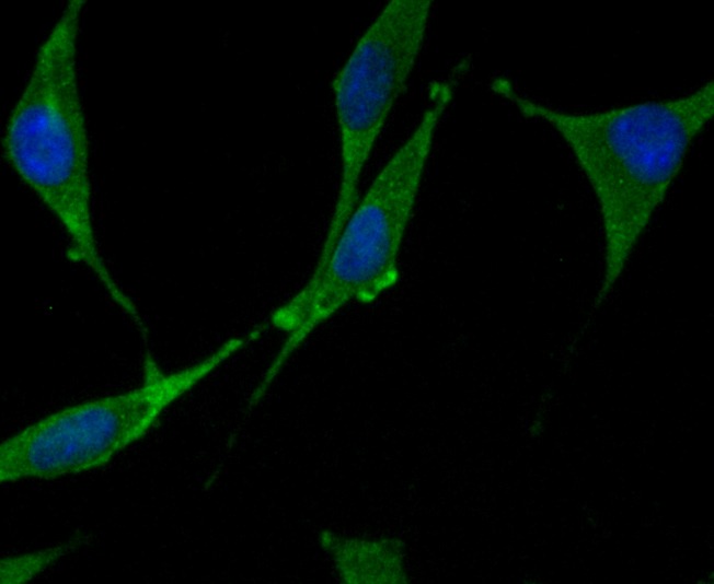 , ,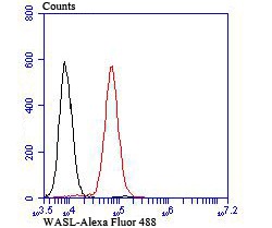 |
| Application | |
| Recommended Dose | WB: 1:500-2000; IHC: 1:50-200; ICC: 1:50-200; FCM: 1:50-100 |
| Antibody Type | Monoclonal |
| Host Species | Rabbit |
| Construction | Recombinant Antibody |
| Purification | ProA affinity purified |
| Appearance | Liquid |
| Formulation | 1*TBS (pH7.4), 1%BSA, 40%Glycerol. Preservative: 0.05% Sodium Azide. |
| Research Background | This gene encodes a member of the Wiskott-Aldrich syndrome (WAS) protein family. Wiskott-Aldrich syndrome proteins share similar domain structure, and associate with a variety of signaling molecules to alter the actin cytoskeleton. The encoded protein is highly expressed in neural tissues, and interacts with several proteins involved in cytoskeletal organization, including cell division control protein 42 (CDC42) and the actin-related protein-2/3 (ARP2/3) complex. The encoded protein may be involved in the formation of long actin microspikes, and in neurite extension. Regulates actin polymerization by stimulating the actin-nucleating activity of the Arp2/3 complex. Involved in mitosis and cytokinesis, via its role in the regulation of actin polymerization. Binds to HSF1/HSTF1 and forms a complex on heat shock promoter elements (HSE) that negatively regulates HSP90 expression.. |
| Conjucates | Unconjugated |
| Immunogen | Recombinant Protein |
| Uniprot ID |
| Molecular Weight | Theoretical: 55 kDa. |
| Stability & Storage | Store at -20°C or -80°C for 12 months. Avoid repeated freeze-thaw cycles. |
| Transport | Shipping with blue ice. |
| Size | Quantity | Unit Price | Amount | Operation |
|---|
 Hello! How can I help you today?
 Copyright © 2015-2026 TargetMol Chemicals Inc. All Rights Reserved.
