
Shopping Cart
Remove All Your shopping cart is currently empty

Synonyms: neurotrophic tyrosine kinase, receptor, type 1
Anti-TrkA Polyclonal Antibody
| Pack Size | Price | USA Stock | Global Stock | Quantity |
|---|---|---|---|---|
| 50 µL | $222 | 7-10 days | 7-10 days | |
| 100 µL | $372 | 7-10 days | 7-10 days | |
| 200 µL | $527 | 7-10 days | 7-10 days |
| Description | Anti-TrkA Polyclonal Antibody is a Rabbit antibody targeting TrkA. Anti-TrkA Polyclonal Antibody can be used in IF, IHC-Fr, IHC-P, WB. |
| Synonyms | neurotrophic tyrosine kinase, receptor, type 1 |
| Ig Type | IgG |
| Reactivity | Human,Mouse,Rat (predicted:Pig,Cow,Horse,Sheep) |
| Verified Activity | 1. Tissue/cell: mouse brain tissue;4% Paraformaldehyde-fixed and paraffin-embedded; Antigen retrieval: citrate buffer (0.01M, pH6.0), Boiling bathing for 15 min; Blocking buffer (normal goat serum) at 37°C for 20 min; Incubation: Anti-TrkA Polyclonal Antibody, Unconjugated (TMAB-01900) 1:200, overnight at 4°C; The secondary antibody was Goat Anti-Rabbit IgG, Cy3 conjugated used at 1:200 dilution for 40 minutes at 37°C. DAPI (5 μg/ml,blue) was used to stain the cell nucleus. 2. Tissue/cell: mouse embryo tissue;4% Paraformaldehyde-fixed and paraffin-embedded; Antigen retrieval: citrate buffer (0.01M, pH6.0), Boiling bathing for 15 min; Blocking buffer (normal goat serum) at 37°C for 20 min; Incubation: Anti-TrkA Polyclonal Antibody, Unconjugated (TMAB-01900) 1:200, overnight at 4°C; The secondary antibody was Goat Anti-Rabbit IgG, Cy3 conjugated used at 1:200 dilution for 40 minutes at 37°C. DAPI (5 μg/ml,blue) was used to stain the cell nucleus. 3. Sample: U251 Cell (Human) Lysate at 40 μg Primary: Anti-TrkA (TMAB-01900) at 1/300 dilution Secondary: IRDye800CW Goat Anti-Rabbit IgG at 1/20000 dilution Predicted band size: 90 kDa Observed band size: 100 kDa 4. Sample: LympHnode (Mouse) Lysate at 40 μg LympHnode (Rat) Lysate at 40 μg Primary: Anti-TrkA (TMAB-01900) at 1/300 dilution Secondary: IRDye800CW Goat Anti-Rabbit IgG at 1/20000 dilution Predicted band size: 90 kDa Observed band size: 100 kDa 5. Paraformaldehyde-fixed, paraffin embedded (Mouse brain); Antigen retrieval by boiling in sodium citrate buffer (pH6.0) for 15 min; Block endogenous peroxidase by 3% hydrogen peroxide for 20 min; Blocking buffer (normal goat serum) at 37°C for 30 min; Antibody incubation with (TrkA) Polyclonal Antibody, Unconjugated (TMAB-01900) at 1:400 overnight at 4°C, followed by operating according to SP Kit (Rabbit) instructionsand DAB staining. 6. Sample: Lane 1: Cerebrum (Mouse) Lysate at 40 μg Primary: Anti-TrkA (TMAB-01900) at 1/1000 dilution Secondary: IRDye800CW Goat Anti-Rabbit IgG at 1/20000 dilution Predicted band size: 130 kDa Observed band size: 130 kDa 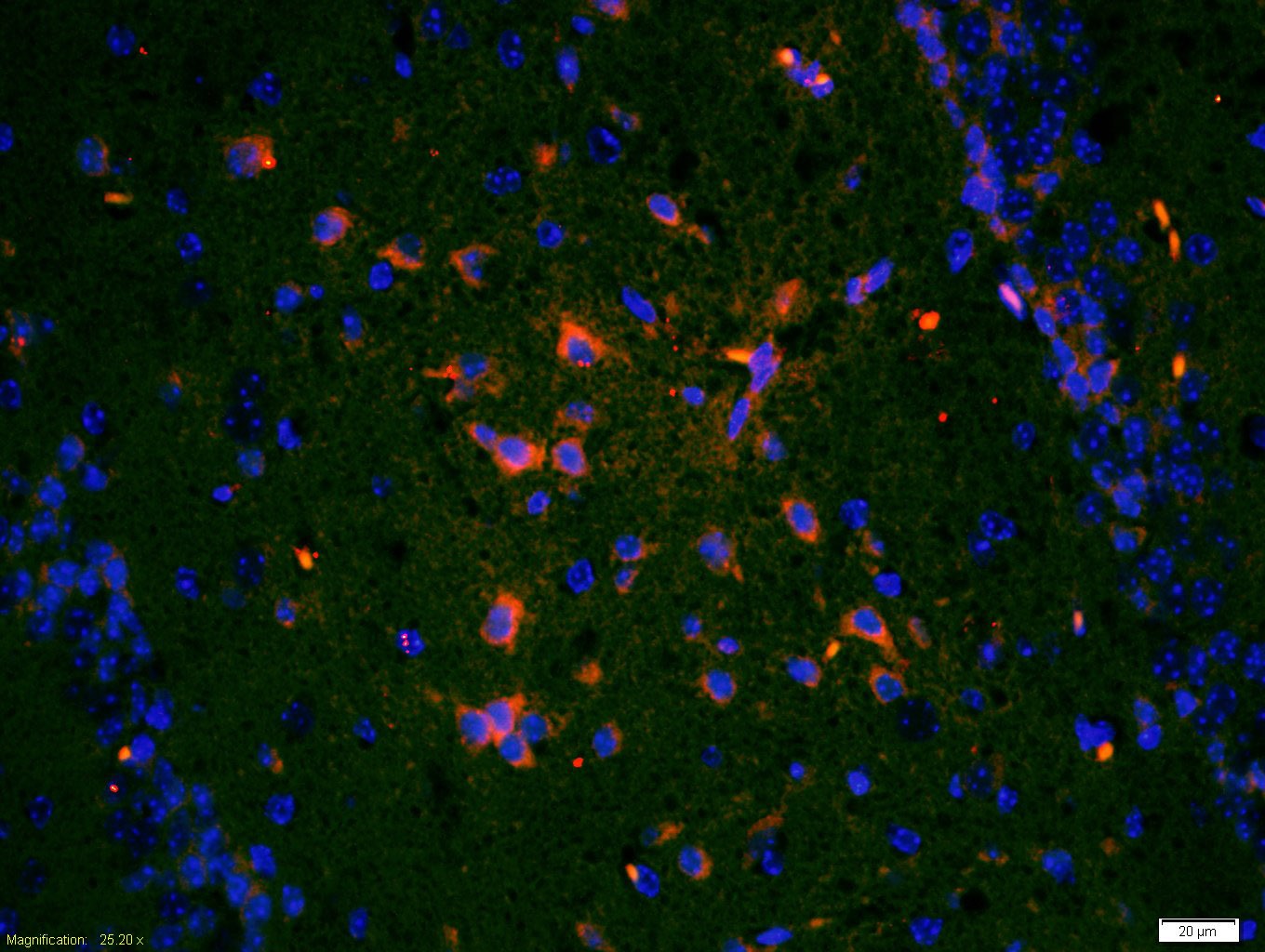 , ,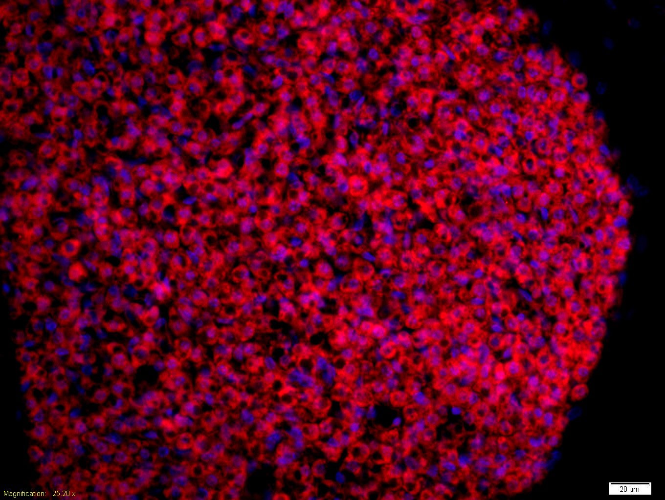 , ,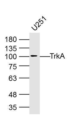 , ,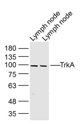 , ,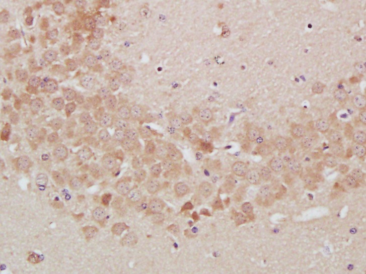 , ,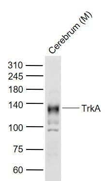 |
| Application | |
| Recommended Dose | WB: 1:500-2000; IHC-P: 1:100-500; IHC-Fr: 1:100-500; IF: 1:100-500 |
| Antibody Type | Polyclonal |
| Host Species | Rabbit |
| Subcellular Localization | Cell membrane; Single-pass type I membrane protein. Early endosome membrane; Single-pass type I membrane protein. Note=Internalized to endosomes upon binding of NGF or NTF3 and further transported to the cell body via a retrograde axonal transport. |
| Tissue Specificity | Isoform TrkA-I is found in most non-neuronal tissues. Isoform TrkA-II is primarily expressed in neuronal cells. TrkA-III is specifically expressed by pluripotent neural stem and neural crest progenitors. |
| Construction | Polyclonal Antibody |
| Purification | Protein A purified |
| Appearance | Liquid |
| Formulation | 0.01M TBS (pH7.4) with 1% BSA, 0.02% Proclin300 and 50% Glycerol. |
| Concentration | 1 mg/mL |
| Research Background | The Trk family of nerve growth factor receptors includes Trk A(also referfed to as Trk A gp140),Trk B and Trk C. The prototype member of this gene family, Trk A, encodes a 140 kDa cell surface receptor , gp140, the expression of which is restricted in vivo to neurons of the sensory spinal and cranial gangliaof neurocrest origin. Nerve growth factor (NGF) stimulates tyrosine phosphorylation of Trk gp 140 in neural cell lines and in embryonic dorsal root ganglia. By comparison, BDNF and to a lesser extent, NT-3, but not NGF, can induce tyrosine phophorylayion of Trk B gp 145. The third member of the Trk receptor family, Trk C incodes a 140 kDa protein, Trk C gp140, that is preferentially expressed in brain tissue and primarily functions as a receptor for NT-3.An additional component of the Trk receptor complex, NGFR p175, binds to neurotrophic factors with low affinity but is required for efficient signaling. NGFR p175 accelerates Trk activation and may recruit downstream dffector molecules to the ligand-bound receptor complex. |
| Immunogen | KLH conjugated synthetic peptide: human Trk A |
| Antigen Species | Human |
| Gene Name | NTRK1 |
| Gene ID | |
| Protein Name | High affinity nerve growth factor receptor |
| Uniprot ID | |
| Function | Receptor tyrosine kinase involved in the development and the maturation of the central and peripheral nervous systems through regulation of proliferation, differentiation and survival of sympathetic and nervous neurons. High affinity receptor for NGF which is its primary ligand, it can also bind and be activated by NTF3/neurotrophin-3. However, NTF3 only supports axonal extension through NTRK1 but has no effect on neuron survival. Upon dimeric NGF ligand-binding, undergoes homodimerization, autophosphorylation and activation. Recruits, phosphorylates and/or activates several downstream effectors including SHC1, FRS2, SH2B1, SH2B2 and PLCG1 that regulate distinct overlapping signaling cascades driving cell survival and differentiation. Through SHC1 and FRS2 activates a GRB2-Ras-MAPK cascade that regulates cell differentiation and survival. Through PLCG1 controls NF-Kappa-B activation and the transcription of genes involved in cell survival. Through SHC1 and SH2B1 controls a Ras-PI3 kinase-AKT1 signaling cascade that is also regulating survival. In absence of ligand and activation, may promote cell death, making the survival of neurons dependent on trophic factors. Isoform TrkA-III is resistant to NGF, constitutively activates AKT1 and NF-kappa-B and is unable to activate the Ras-MAPK signaling cascade. Antagonizes the anti-proliferative NGF-NTRK1 signaling that promotes neuronal precursors differentiation. Isoform TrkA-III promotes angiogenesis and has oncogenic activity when overexpressed. |
| Molecular Weight | Theoretical: 90 kDa. Actual: 100 kDa. |
| Stability & Storage | Store at -20°C or -80°C for 12 months. Avoid repeated freeze-thaw cycles. |
| Transport | Shipping with blue ice. |
| Size | Quantity | Unit Price | Amount | Operation |
|---|
 Hello! How can I help you today?
 Copyright © 2015-2026 TargetMol Chemicals Inc. All Rights Reserved.
