Shopping Cart
Remove All Your shopping cart is currently empty
Anti-Syncytin-1 Polyclonal Antibody is a Rabbit antibody targeting Syncytin-1. Anti-Syncytin-1 Polyclonal Antibody can be used in WB.
| Pack Size | Price | USA Warehouse | Global Warehouse | Quantity |
|---|---|---|---|---|
| 50 μL | $221 | 7-10 days | 7-10 days | |
| 100 μL | $372 | 7-10 days | 7-10 days | |
| 200 μL | $528 | 7-10 days | 7-10 days |
| Description | Anti-Syncytin-1 Polyclonal Antibody is a Rabbit antibody targeting Syncytin-1. Anti-Syncytin-1 Polyclonal Antibody can be used in WB. |
| Synonyms | Syncytin-1, Syncytin, HERV-W_7q21.2 provirus ancestral Env polyprotein, HERV-W envelope protein, HERV-7q Envelope protein, ERVWE1, ERVW-1, Env-W, Enverin, Envelope polyprotein gPr73, Endogenous retrovirus group W member 1 |
| Ig Type | IgG |
| Reactivity | Human,Mouse |
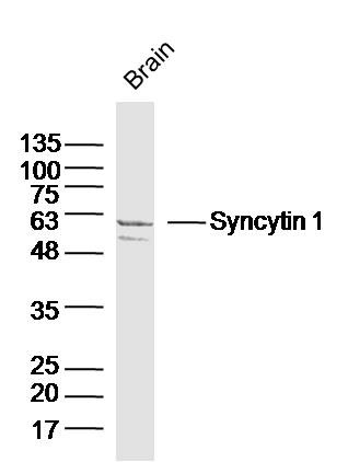
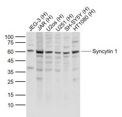
| Verified Activity | 1. Sample: Brain (Mouse) Lysate at 40 μg Primary: Anti-Syncytin 1 (TMAB-01803) at 1/300 dilution Secondary: IRDye800CW Goat Anti-Rabbit IgG at 1/20000 dilution Predicted band size: 33/58 kDa Observed band size: 58 kDa 2. Sample: Lane 1: JEG-3 (Human) Cell Lysate at 30 μg Lane 2: JAR (Human) Cell Lysate at 30 μg Lane 3: U2os (Human) Cell Lysate at 30 μg Lane 4: U251 (Human) Cell Lysate at 30 μg Lane 5: SH-SY5Y (Human) Cell Lysate at 30 μg Lane 6: HT1080 (Human) Cell Lysate at 30 μg Primary: Anti-Syncytin 1 (TMAB-01803) at 1/1000 dilution Secondary: IRDye800CW Goat Anti-Rabbit IgG at 1/20000 dilution Predicted band size: 58 kDa Observed band size: 60 kDa   |
| Application | |
| Recommended Dose | WB: 1:500-2000 |
| Antibody Type | Polyclonal |
| Host Species | Rabbit |
| Subcellular Localization | Cell membrane |
| Tissue Specificity | Expressed at higher level in placental syncytiotrophoblast. Expressed at intermediate level in testis. Seems also to be found at low level in adrenal tissue, bone marrow, breast, colon, kidney, ovary, prostate, skin, spleen, thymus, thyroid, brain and tra |
| Construction | Polyclonal Antibody |
| Purification | Protein A purified |
| Appearance | Liquid |
| Formulation | 0.01M TBS (pH7.4) with 1% BSA, 0.02% Proclin300 and 50% Glycerol. |
| Concentration | 1 mg/mL |
| Research Background | Many different human endogenous retrovirus (HERV) families are expressed in normal placental tissue at high levels, suggesting that HERVs are functionally important in reproduction. This gene is part of an HERV provirus on chromosome 7 that has inactivating mutations in the gag and pol genes. This gene is the envelope glycoprotein gene which appears to have been selectively preserved. The gene's protein product is expressed in the placental syncytiotrophoblast and is involved in fusion of the cytotrophoblast cells to form the syncytial layer of the placenta. The protein has the characteristics of a typical retroviral envelope protein, including a furin cleavage site that separates the surface (SU) and transmembrane (TM) proteins which form a heterodimer. Alternatively spliced transcript variants encoding the same protein have been found for this gene. [provided by RefSeq, Mar 2010] |
| Immunogen | KLH conjugated synthetic peptide: human Syncytin 1 |
| Antigen Species | Human |
| Gene Name | ERVW-1 |
| Gene ID | |
| Protein Name | Endogenous retrovirus group W member 1 |
| Uniprot ID | |
| Function | This endogenous retroviral envelope protein has retained its original fusogenic properties and participates in trophoblast fusion and the formation of a syncytium during placenta morphogenesis. May induce fusion through binding of SLC1A4 and SLC1A5 (PubMed:10708449, PubMed:12050356, PubMed:23492904).3 PublicationsEndogenous envelope proteins may have kept, lost or modified their original function during evolution. Retroviral envelope proteins mediate receptor recognition and membrane fusion during early infection. The surface protein (SU) mediates receptor recognition, while the transmembrane protein (TM) acts as a class I viral fusion protein. The protein may have at least 3 conformational states: pre-fusion native state, pre-hairpin intermediate state, and post-fusion hairpin state. During viral and target cell membrane fusion, the coiled coil regions (heptad repeats) assume a trimer-of-hairpins structure, positioning the fusion peptide in close proximity to the C-terminal region of the ectodomain. The formation of this structure appears to drive apposition and subsequent fusion of membranes. |
| Molecular Weight | Theoretical: 59 kDa. |
| Stability & Storage | Store at -20°C or -80°C for 12 months. Avoid repeated freeze-thaw cycles. |
| Transport | Shipping with blue ice. |
| Size | Quantity | Unit Price | Amount | Operation |
|---|

Copyright © 2015-2026 TargetMol Chemicals Inc. All Rights Reserved.