
Shopping Cart
Remove All Your shopping cart is currently empty

Synonyms: SLF, Sl, SF, SCF, Mgf, Kitlg, KIT ligand, Gb, Con, Clo
Anti-SCF Polyclonal Antibody
| Pack Size | Price | USA Stock | Global Stock | Quantity |
|---|---|---|---|---|
| 50 µL | $222 | 7-10 days | 7-10 days | |
| 100 µL | $373 | 7-10 days | 7-10 days | |
| 200 µL | $529 | 7-10 days | 7-10 days |
| Description | Anti-SCF Polyclonal Antibody is a Rabbit antibody targeting SCF. Anti-SCF Polyclonal Antibody can be used in FCM,IF,IHC-Fr,IHC-P,WB. |
| Synonyms | SLF, Sl, SF, SCF, Mgf, Kitlg, KIT ligand, Gb, Con, Clo |
| Ig Type | IgG |
| Reactivity | Human,Mouse,Rat (predicted:Goat) |
| Verified Activity | 1. Blank control (Black line): U87MG (Black). Primary Antibody (green line): Rabbit Anti-SCF antibody (TMAB-01667) Dilution: 1 μg/10^6 cells; Isotype Control Antibody (orange line): Rabbit IgG. Secondary Antibody (white blue line): Goat anti-rabbit IgG-PE Dilution: 1 μg/test. Protocol The cells were fixed with 4% PFA (10 min at room temperature) and then were incubated in 5% BSA to block non-specific protein-protein interactions for 30 min at room temperature. Cells stained with Primary Antibody for 30 min at room temperature. The secondary antibody used for 40 min at room temperature. 2. Sample: Lane 1: Lung (Rat) Lysate at 40 μg Lane 2: Spleen (Rat) Lysate at 40 μg Lane 3: MCF-7 (Human) Cell Lysate at 30 μg Lane 4: Hela (Human) Cell Lysate at 30 μg Primary: Anti-SCF (TMAB-01667) at 1/1000 dilution Secondary: IRDye800CW Goat Anti-Rabbit IgG at 1/20000 dilution Predicted band size: 31 kDa Observed band size: 45/35 kDa 3. Paraformaldehyde-fixed, paraffin embedded (Rat brain); Antigen retrieval by boiling in sodium citrate buffer (pH6.0) for 15 min; Block endogenous peroxidase by 3% hydrogen peroxide for 20 min; Blocking buffer (normal goat serum) at 37°C for 30 min; Antibody incubation with (SCF) Polyclonal Antibody, Unconjugated (TMAB-01667) at 1:400 overnight at 4°C, followed by operating according to SP Kit (Rabbit) instructions and DAB staining. 4. Blank control (Black line): Mouse spleen (Black). Primary Antibody (green line): Rabbit Anti-SCF antibody (TMAB-01667) Dilution: 3 μg/10^6 cells; Isotype Control Antibody (orange line): Rabbit IgG. Secondary Antibody (white blue line): Goat anti-rabbit IgG-AF647 Dilution: 1 μg/test. Protocol The cells were fixed with 4% PFA (10 min at room temperature) and then permeabilized with 90% ice-cold methanol for 20 min at room temperature. The cells were then incubated in 5% BSA to block non-specific protein-protein interactions for 30 min at room temperature. Cells stained with Primary Antibody for 30 min at room temperature. The secondary antibody used for 40 min at room temperature. Acquisition of 10,000 events was performed. 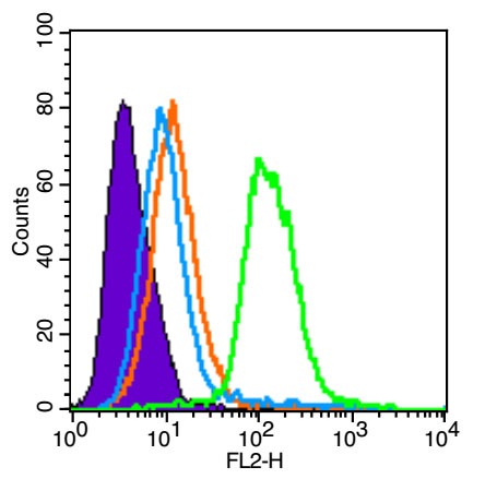 , ,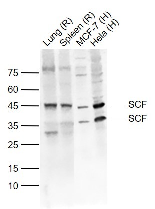 , ,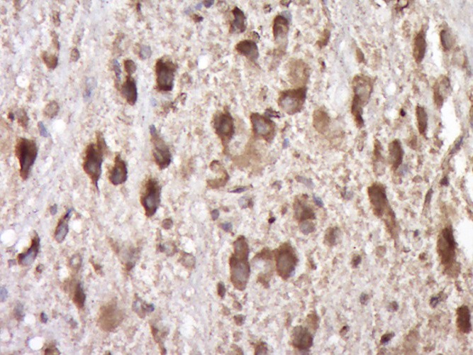 , ,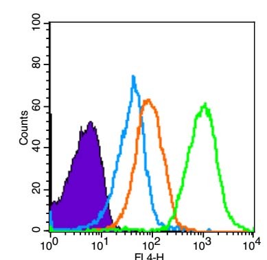 |
| Application | |
| Recommended Dose | WB: 1:500-2000; IHC-P: 1:100-500; IHC-Fr: 1:100-500; IF: 1:100-500; FCM: 1ug/Test |
| Antibody Type | Polyclonal |
| Host Species | Rabbit |
| Subcellular Localization | Isoform 1: Cell membrane; Single-pass type I membrane protein. Isoform 2: Cell membrane; Single-pass type I membrane protein. Cytoplasm, cytoskeleton. Soluble KIT ligand: Secreted. |
| Construction | Polyclonal Antibody |
| Purification | Protein A purified |
| Appearance | Liquid |
| Formulation | 0.01M TBS (pH7.4) with 1% BSA, 0.02% Proclin300 and 50% Glycerol. |
| Concentration | 1 mg/mL |
| Research Background | Stem Cell Factor (SCF), also known as c-Kit ligand (KL), steel factor (SLF) and mast cell growth factor (MGF), is a 30 kDa glycoprotein with broad activities on various tissues, including hematopoietic cells, pigment cells, and primordial germ cells. SCF is secreted by endothelial cells, fibroblasts, and bone marrow stromal cells as a membrane-bound form which may be cleaved to release the soluble form. Both forms are active in promoting colony formation from murine bone marrow cells, but membrane-bound SCF is more effective in promoting hematopoieses in vivo, suggesting a role in cellular interactions between hematopoietic and stromal cells. The soluble form is thought to exist in solution as a noncovalently linked dimer. SCF is structurally related to M-CSF (CSF-1) and Flt-3/Flk-2 Ligand (FL) with all three sharing a similar size, existence of transmembrane and soluble forms, four conserved cysteines, and alternative splicing exon locations, but they share little sequence homology. SCF alone is a modest colony stimulating factor. However, in the presence of other cytokines such as EPO, TPO, GM-CSF, G-CSF, M-CSF, IL-3, and IL-7, SCF is a potent costimulant that works synergistically to increase the size of myeloid, erythroid or lymphoid lineage colonies without influencing the lineage differentiation of the progenitors. |
| Immunogen | KLH conjugated synthetic peptide: human SCF |
| Antigen Species | Human |
| Gene Name | KITLG |
| Gene ID | |
| Protein Name | Kit ligand |
| Uniprot ID | |
| Biology Area | Surface molecules,CSFs,Surface Molecules |
| Molecular Weight | Theoretical: 31 kDa. Actual: 45/35 kDa. |
| Stability & Storage | Store at -20°C or -80°C for 12 months. Avoid repeated freeze-thaw cycles. |
| Transport | Shipping with blue ice. |
| Size | Quantity | Unit Price | Amount | Operation |
|---|
 Hello! How can I help you today?
 Copyright © 2015-2026 TargetMol Chemicals Inc. All Rights Reserved.
