
Shopping Cart
Remove All Your shopping cart is currently empty

Synonyms:
Anti-PRDX3 Antibody
(7V569)
| Pack Size | Price | USA Stock | Global Stock | Quantity |
|---|---|---|---|---|
| 50 µL | $297 | 7-10 days | 7-10 days | |
| 100 µL | $498 | 7-10 days | 7-10 days |
| Description | Anti-PRDX3 Antibody (7V569) is a Rabbit antibody targeting PRDX3. Anti-PRDX3 Antibody (7V569) can be used in FCM,ICC,IHC,IP,WB. |
| Ig Type | IgG |
| Clone | 7V569 |
| Reactivity | Human |
| Verified Activity | 1. Western blot analysis of Peroxiredoxin 3 on different cell lysate using anti-Peroxiredoxin 3 antibody at 1/1,000 dilution. Positive control: Lane1: Human liver, Lane2: MCF-7, Lane3: A431. 2. Immunohistochemical analysis of paraffin-embedded human liver tissue using anti- Peroxiredoxin 3 antibody. Counter stained with hematoxylin. 3. Immunohistochemical analysis of paraffin-embedded human liver cancer tissue using anti- Peroxiredoxin 3 antibody. Counter stained with hematoxylin. 4. Immunohistochemical analysis of paraffin-embedded human kidney tissue using anti- Peroxiredoxin 3 antibody. Counter stained with hematoxylin. 5. ICC staining Peroxiredoxin 3 in Hela cells (green). The nuclear counter stain is DAPI (blue). Cells were fixed in paraformaldehyde, permeabilised with 0.25% Triton X100/PBS. 6. ICC staining Peroxiredoxin 3 in HepG2 cells (green). The nuclear counter stain is DAPI (blue). Cells were fixed in paraformaldehyde, permeabilised with 0.25% Triton X100/PBS. 7. ICC staining Peroxiredoxin 3 in MCF-7 cells (green). The nuclear counter stain is DAPI (blue). Cells were fixed in paraformaldehyde, permeabilised with 0.25% Triton X100/PBS. 8. Flow cytometric analysis of MCF-7 cells with Peroxiredoxin 3 antibody at 1/100 dilution (red) compared with an unlabelled control (cells without incubation with primary antibody; black). 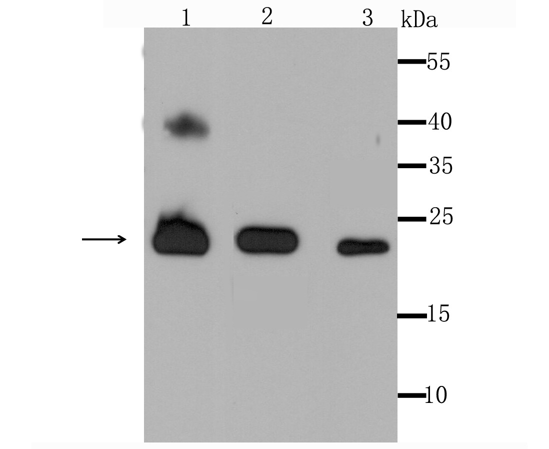 , ,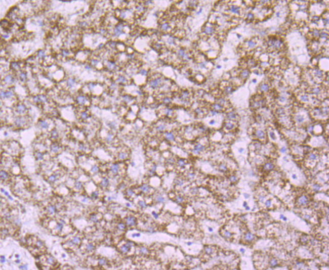 , ,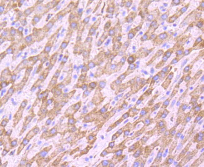 , ,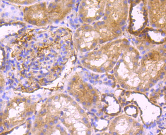 , ,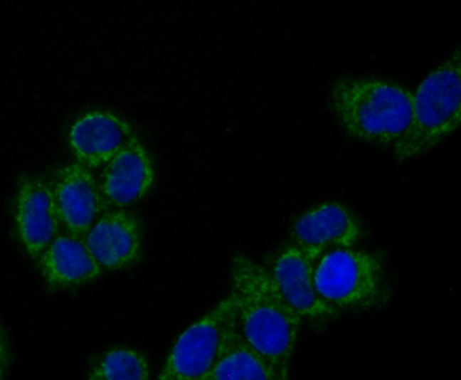 , ,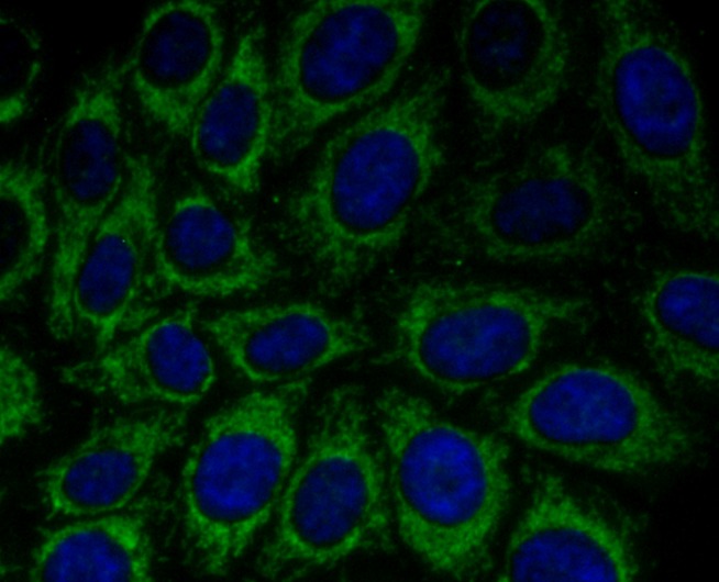 , ,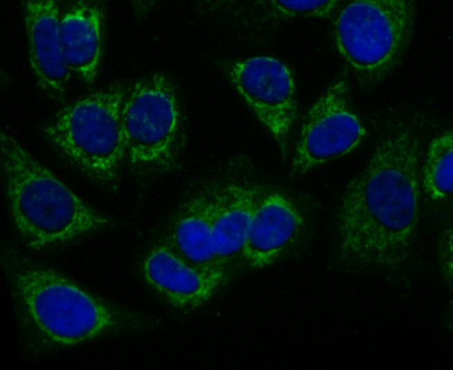 , ,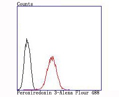 |
| Application | |
| Recommended Dose | WB: 1:500-2000; IHC: 1:50-200; ICC: 1:50-200; FCM: 1:50-100 |
| Antibody Type | Monoclonal |
| Host Species | Rabbit |
| Construction | Recombinant Antibody |
| Purification | ProA affinity purified |
| Appearance | Liquid |
| Formulation | 1*TBS (pH7.4), 1%BSA, 40%Glycerol. Preservative: 0.05% Sodium Azide. |
| Research Background | The peroxiredoxin (PRX) family comprises six antioxidant proteins, PRX I, II, III, IV, V and VI, which protect cells from reactive oxygen species (ROS) by preventing the metal-catalyzed oxidation of enzymes. The PRX proteins primarily utilize thioredoxin as the electron donor for antioxidation, although they are fairly promiscuous with regard to the hydroperoxide substrate. In addition to protection from ROS, peroxiredoxins are also involved in cell proliferation, differentiation and gene expression. PRX I, II, IV and VI show diffuse cytoplasmic localization, while PRX III and V exhibit distinct mitochondrial localization. The human PRX I gene encodes a protein that is expressed in several tissues, including liver, kidney, testis, lung and nervous system. PRX II is expressed in testis, while PRX III shows expression in lung. PRX I, II and III are overexpressed in breast cancer and may be involved in its development or progression. Upregulated protein levels of PRX I and II in Alzheimer's disease (AD) and Down syndrome (DS) indicate the involvement of PRX I and II in their pathogenesis. The human PRX IV gene is abundantly expressed in many tissues. |
| Conjucates | Unconjugated |
| Immunogen | Recombinant Protein |
| Uniprot ID |
| Molecular Weight | Theoretical: 24 kDa. |
| Stability & Storage | Store at -20°C or -80°C for 12 months. Avoid repeated freeze-thaw cycles. |
| Transport | Shipping with blue ice. |
| Size | Quantity | Unit Price | Amount | Operation |
|---|
 Hello! How can I help you today?
 Copyright © 2015-2026 TargetMol Chemicals Inc. All Rights Reserved.
