
Shopping Cart
Remove All Your shopping cart is currently empty

Synonyms: p-MLKL (Ser345), p-MLKL (S345), MLKL (p-Ser345), MLKL (p-S345), Mlkl, Mixed lineage kinase domain-like protein
Anti-Phospho-MLKL
(Ser345) Antibody
(2S387)
| Pack Size | Price | USA Stock | Global Stock | Quantity |
|---|---|---|---|---|
| 25 µL | $153 | 7-10 days | 7-10 days | |
| 50 µL | $272 | 7-10 days | 7-10 days | |
| 100 µL | $487 | 7-10 days | 7-10 days |
| Description | Anti-Phospho-MLKL (Ser345) Antibody (2S387) is a Rabbit antibody targeting HLA F. Anti-Phospho-MLKL (Ser345) Antibody (2S387) can be used in WB,ICC/IF. |
| Synonyms | p-MLKL (Ser345), p-MLKL (S345), MLKL (p-Ser345), MLKL (p-S345), Mlkl, Mixed lineage kinase domain-like protein |
| Ig Type | IgG |
| Clone | 2S387 |
| Reactivity | Mouse |
| Verified Activity | 1. Western blot analysis of Phospho-MLKL (S345) on L929 cell lysates. Lane 1: L929 cells, whole cell lysate, 10 μg/lane. Lane 2: L929 cells were treated with 20 uM Z-VAD for 30 minutes, then added 20 ng/ml mTNF-alpha and 100 nM SM-164 for 4 hours, whole cell lysates, 10 μg/lane. 2. 4% Paraformaldehyde-fixed L-929 (treated with 20 ng/ml TNF alpha, 100 nM Smac mimetic, and 20 µM z-VAD for 8 h) (M) cell; Triton X-100 at RT for 20 min; Antibody incubation with (phospho-MLKL (Ser345)) monoclonal Antibody, unconjugated (TMAB-01454) 1:100, 90 min at 37°C; followed by conjugated Goat Anti-Rabbit IgG antibody (green) at 37°C for 90 min, DAPI (blue) was used to stain the cell nucleus. PBS instead of the primary antibody was used as the blank control. 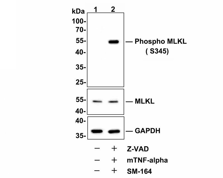 , ,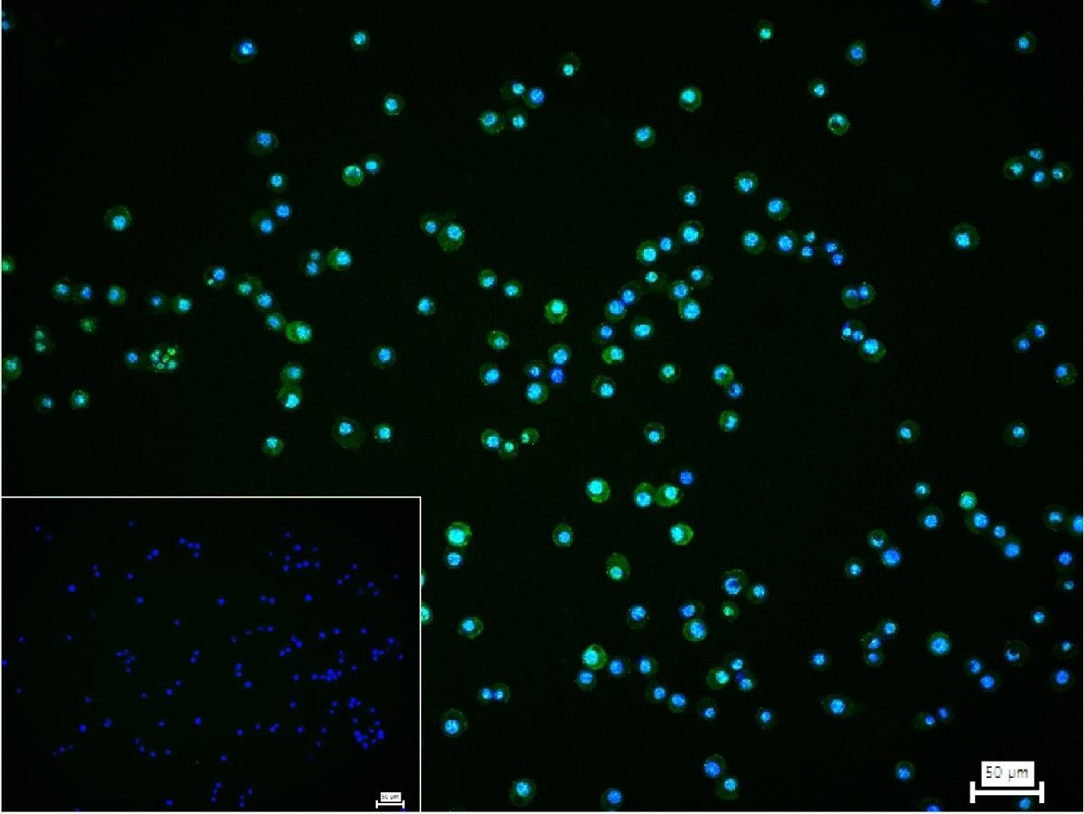 |
| Application | |
| Recommended Dose | WB=1:500-2000,ICC/IF=1:50-200 |
| Antibody Type | Monoclonal |
| Host Species | Rabbit |
| Subcellular Localization | Cytoplasm. Cell membrane. Note=Localizes to the cytoplasm and translocates to the plasma membrane on necroptosis induction. |
| Construction | Recombinant Antibody |
| Purification | Protein A purified |
| Appearance | Liquid |
| Formulation | 0.01M TBS (pH7.4) with 1% BSA, 0.02% Proclin300 and 50% Glycerol. |
| Concentration | 1 mg/mL |
| Research Background | This gene belongs to the protein kinase superfamily. The encoded protein contains a protein kinase-like domain; however, is thought to be inactive because it lacks several residues required for activity. This protein plays a critical role in tumor necrosis factor (TNF)-induced necroptosis, a programmed cell death process, via interaction with receptor-interacting protein 3 (RIP3), which is a key signaling molecule in necroptosis pathway. Inhibitor studies and knockdown of this gene inhibited TNF-induced necrosis. High levels of this protein and RIP3 are associated with inflammatory bowel disease in children. Alternatively spliced transcript variants have been described for this gene. [provided by RefSeq, Sep 2015]. |
| Immunogen | A synthesized peptide: mouse Mlkl around the phosphorylation site of S345 |
| Antigen Species | Mouse |
| Gene Name | MLKL |
| Gene ID | |
| Protein Name | Mixed lineage kinase domain-like protein |
| Uniprot ID | |
| Biology Area | Necroptosis,SARS Coronavirus,Other Kinases |
| Function | Pseudokinase that plays a key role in TNF-induced necroptosis, a programmed cell death process. Activated following phosphorylation by RIPK3, leading to homotrimerization, localization to the plasma membrane and execution of programmed necrosis characterized by calcium influx and plasma membrane damage. Does not have protein kinase activity. |
| Molecular Weight | Theoretical: 54 kDa. Actual: 54 kDa. |
| Stability & Storage | Store at -20°C or -80°C for 12 months. Avoid repeated freeze-thaw cycles. |
| Transport | Shipping with blue ice. |
| Size | Quantity | Unit Price | Amount | Operation |
|---|
 Hello! How can I help you today?
 Copyright © 2015-2026 TargetMol Chemicals Inc. All Rights Reserved.
