
Shopping Cart
Remove All Your shopping cart is currently empty

Synonyms: Phospho-Histone H1.4 (T17), p-Histone H1.4 (Thr17), p-Histone H1.4 (T17), MGC116819, Histone H1B, Histone H1.4 (p-Thr17), Histone H1.4 (p-T17), Histone H1, Histone cluster 1 H1e, Histone 1 H1e, Hist1h1e, H1F4, H1E, H14_HUMAN, H1.4, H1 histone family member 4
Anti-Phospho-Histone H1.3/1.4
(Thr17) Antibody
(6O691)
| Pack Size | Price | USA Stock | Global Stock | Quantity |
|---|---|---|---|---|
| 50 µL | $298 | 7-10 days | 7-10 days | |
| 100 µL | $496 | 7-10 days | 7-10 days |
| Description | Anti-Phospho-Histone H1.3/1.4 (Thr17) Antibody (6O691) is a Rabbit antibody targeting Phospho-Histone H1.3/1.4 (Thr17). Anti-Phospho-Histone H1.3/1.4 (Thr17) Antibody (6O691) can be used in ICC/IF,IHC,WB. |
| Synonyms | Phospho-Histone H1.4 (T17), p-Histone H1.4 (Thr17), p-Histone H1.4 (T17), MGC116819, Histone H1B, Histone H1.4 (p-Thr17), Histone H1.4 (p-T17), Histone H1, Histone cluster 1 H1e, Histone 1 H1e, Hist1h1e, H1F4, H1E, H14_HUMAN, H1.4, H1 histone family member 4 |
| Ig Type | IgG |
| Clone | 6O691 |
| Reactivity | Human,Mouse,Rat |
| Verified Activity | 1. Western blot analysis of Phospho-Histone H1.3 (T17)+Histone H1.4 (T17) on CRC cell lysates using anti-Phospho-Histone H1.3 (T17)+Histone H1.4 (T17) antibody at 1/500 dilution. Positive control: Lane 1: Untreated CRC whole cell lysates, Lane2: CRC cells treated with 1.5ug/ml Colcemid for 12 hours whole cell lysates. 2. Immunohistochemical analysis of paraffin-embedded human colon cancer tissue using anti-Phospho-Histone H1.3 (T17)+Histone H1.4 (T17) antibody. Counter stained with hematoxylin. 3. Immunohistochemical analysis of paraffin-embedded mouse colon cancer tissue using anti-Phospho-Histone H1.3 (T17)+Histone H1.4 (T17) antibody. Counter stained with hematoxylin. 4. ICC staining Phospho-Histone H1.3 (T17)+Histone H1.4 (T17) in NIH/3T3 cells (green). The nuclear counter stain is DAPI (blue). Cells were fixed in paraformaldehyde, permeabilised with 0.25% Triton X100/PBS. 5. ICC staining Phospho-Histone H1.3 (T17)+Histone H1.4 (T17) in CRC cells (green). Cells were fixed in paraformaldehyde, permeabilised with 0.25% Triton X100/PBS. 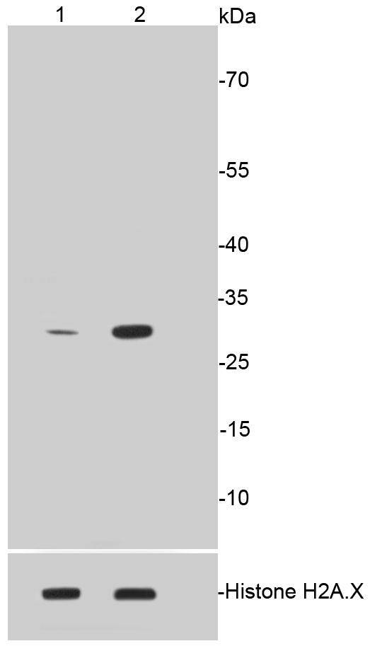 , ,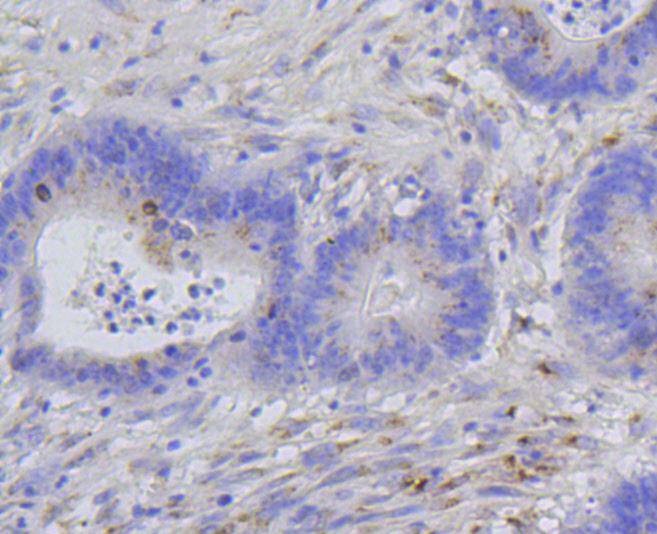 , ,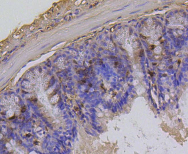 , ,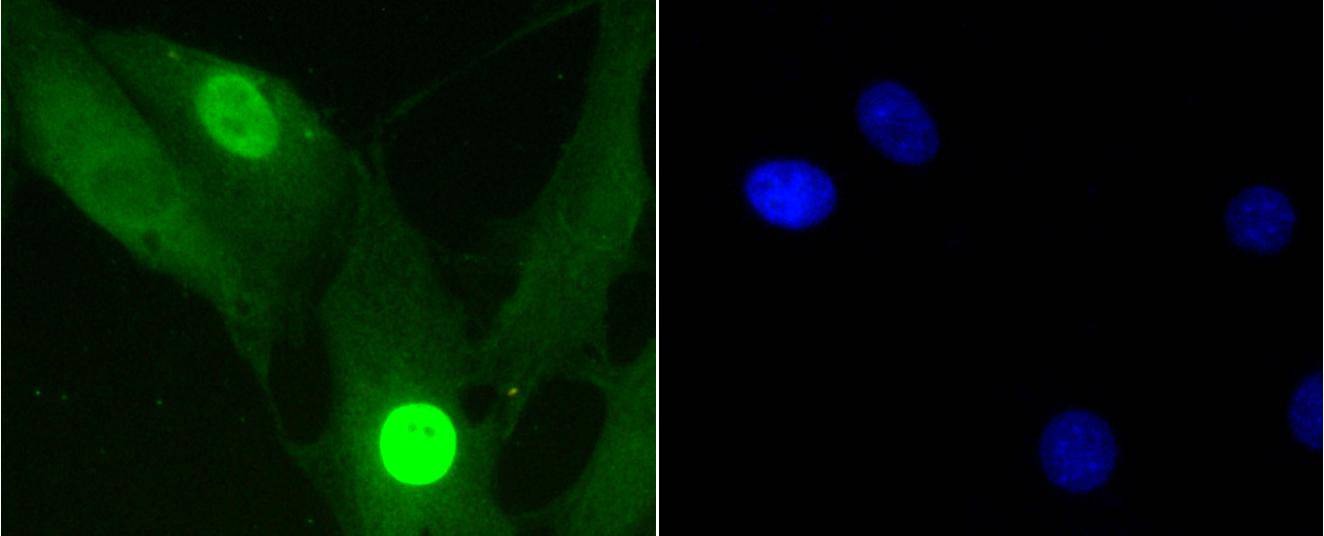 , ,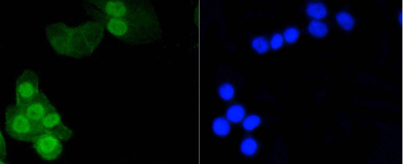 |
| Application | |
| Recommended Dose | WB: 1:500-1000; IHC: 1:50-200; ICC/IF: 1:50-200 |
| Antibody Type | Monoclonal |
| Host Species | Rabbit |
| Construction | Recombinant Antibody |
| Purification | ProA affinity purified |
| Appearance | Liquid |
| Formulation | 1*TBS (pH7.4), 1%BSA, 40%Glycerol. Preservative: 0.05% Sodium Azide. |
| Research Background | Eukaryotic histones are basic and water soluble nuclear proteins that form hetero-octameric nucleosome particles by wrapping 146 base pairs of DNA in a left-handed super-helical turn sequentially to form chromosomal fiber. Two molecules of each of the four core histones (H2A, H2B, H3, and H4) form the octamer; formed of two H2A-H2B dimers and two H3-H4 dimers, forming two nearly symmetrical halves by tertiary structure. Over 80% of nucleosomes contain the linker Histone H1, derived from an intronless gene, that interacts with linker DNA between nucleosomes and mediates compaction into higher order chromatin. Histones are subject to posttranslational modification by enzymes primarily on their N-terminal tails, but also in their globular domains. Such modifications include methylation, citrullination, acetylation, phosphorylation, sumoylation, ubiquitination and ADP-ribosylation. |
| Conjucates | Unconjugated |
| Others Formats | Phospho |
| Immunogen | A synthesized phosphopeptide: human H1.4 around the phosphorylation site of Thr17 |
| Antigen Species | Human |
| Molecular Weight | Theoretical: 30 kDa. |
| Stability & Storage | Store at -20°C or -80°C for 12 months. Avoid repeated freeze-thaw cycles. |
| Transport | Shipping with blue ice. |
| Size | Quantity | Unit Price | Amount | Operation |
|---|
 Hello! How can I help you today?
 Copyright © 2015-2026 TargetMol Chemicals Inc. All Rights Reserved.
