
Shopping Cart
Remove All Your shopping cart is currently empty

Synonyms: Phospho-GSK3B (S9), p-GSK3B (Ser9), p-GSK3B (S9), GSK-3β, GSK3β, GSK3B (p-Ser9), GSK3B (p-S9), glycogen synthase kinase 3 β, glycogen synthase kinase 3 beta
Anti-Phospho-GSK3B
(Ser9) Polyclonal Antibody
| Pack Size | Price | USA Stock | Global Stock | Quantity |
|---|---|---|---|---|
| 50 µL | $222 | 7-10 days | 7-10 days | |
| 100 µL | $374 | 7-10 days | 7-10 days | |
| 200 µL | $527 | 7-10 days | 7-10 days |
| Description | Anti-Phospho-GSK3B (Ser9) Polyclonal Antibody is a Rabbit antibody targeting Phospho-GSK3B (Ser9). Anti-Phospho-GSK3B (Ser9) Polyclonal Antibody can be used in FCM, ICC/IF, IF, IHC-Fr, IHC-P, WB. |
| Synonyms | Phospho-GSK3B (S9), p-GSK3B (Ser9), p-GSK3B (S9), GSK-3β, GSK3β, GSK3B (p-Ser9), GSK3B (p-S9), glycogen synthase kinase 3 β, glycogen synthase kinase 3 beta |
| Ig Type | IgG |
| Reactivity | Human,Mouse,Rat |
| Verified Activity | 1. Sample: Testis (Rat) Lysate at 30 μg Primary: Anti-phospho-GSK-3 Beta (Ser9) (TMAB-01416) at 1:200 dilution; Secondary: HRP conjugated Goat Anti-Rabbit Igg (secondary antibody) at 1: 5000 dilution; Predicted band size: 47 kDa Observed band size: 49 kDa 2. The blue histogram is unstained cells (A549 cells). The Orange histogram is cells stained with Rabbit IgG/FITC. The green histogram is cells stained with Rabbit Anti-phospho-GSK-3 Beta (Ser9)/FITC Conjugated antibody (TMAB-01416-FITC). Isotype control: Cell lines treated with Rabbit IgG/FITC instead of the primary antibody to confirm that primary antibody binding is specific. Concentration: 5 μL in 100 μL 1 X PBS containing 0.5% BSA. 3. Tissue/cell: rat kidney tissue; 4% Paraformaldehyde-fixed and paraffin-embedded; Antigen retrieval: citrate buffer (0.01M, pH6.0), Boiling bathing for 15 min; Block endogenous peroxidase by 3% Hydrogen peroxide for 30 min; Blocking buffer (normal goat serum) at 37°C for 20 min; Incubation: Anti-phospho-GSK-3 Beta (Ser9) Polyclonal Antibody, Unconjugated (TMAB-01416) 1:200, overnight at 4°C, followed by conjugation to the secondary antibody and DAb staining. 4. Paraformaldehyde-fixed, paraffin embedded (Mouse placenta); Antigen retrieval by boiling in sodium citrate buffer (pH6.0) for 15 min; Block endogenous peroxidase by 3% hydrogen peroxide for 20 min; Blocking buffer (normal goat serum) at 37°C for 30 min; Antibody incubation with (Beta (Ser9)) Polyclonal Antibody, Unconjugated (TMAB-01416 p-GSK-3) at 1:500 overnight at 4°C, followed by a conjugated secondary for 20 min and DAB staining. 5. Blank control: A431. Primary Antibody (green line): Rabbit Anti-phospho-GSK-3 Beta (Ser9) antibody (TMAB-01416) Dilution: 1 μg/10^6 cells; Isotype Control Antibody (orange line): Rabbit IgG. Secondary Antibody: Goat anti-rabbit IgG-AF488 Dilution: 1 μg/test. Protocol The cells were fixed with 4% PFA (10 min at room temperature) and then permeabilized with 90% ice-cold methanol for 20 min at-20°C. The cells were then incubated in 5% BSA to block non-specific protein-protein interactions for 30 min at room temperature. Cells stained with Primary Antibody for 30 min at room temperature. The secondary antibody used for 40 min at room temperature. 6. Tissue/cell: Hela cell; 4% Paraformaldehyde-fixed; Triton X-100 at room temperature for 20 min; Blocking buffer (normal goat serum) at 37°C for 20 min; Antibody incubation with (phospho-GSK-3 Beta (Ser9)) polyclonal Antibody, Unconjugated (TMAB-01416) 1:100, 90 minutes at 37°C; followed by a FITC conjugated Goat Anti-Rabbit IgG antibody at 37°C for 90 minutes, DAPI (blue) was used to stain the cell nucleus. 7. Sample: Lane 1: Mouse Cerebrum tissue lysates Lane 2: Rat Cerebrum tissue lysates Primary: Anti-phospho-GSK-3 Beta (Ser9) (TMAB-01416) at 1/1000 dilution Secondary: IRDye800CW Goat Anti-Rabbit IgG at 1/20000 dilution Predicted band size: 47 kDa Observed band size: 47 kDa 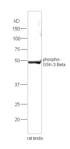 , ,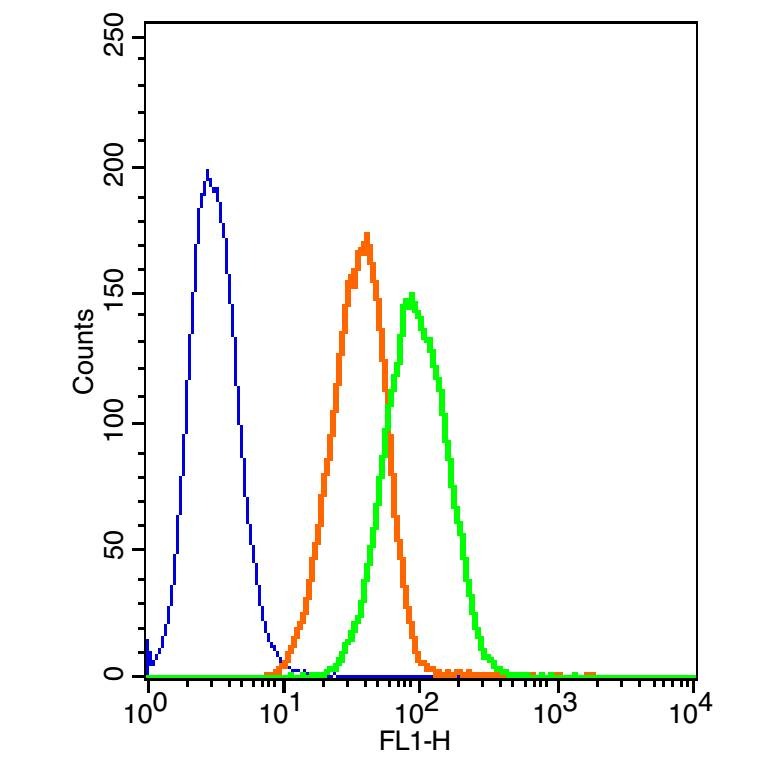 , ,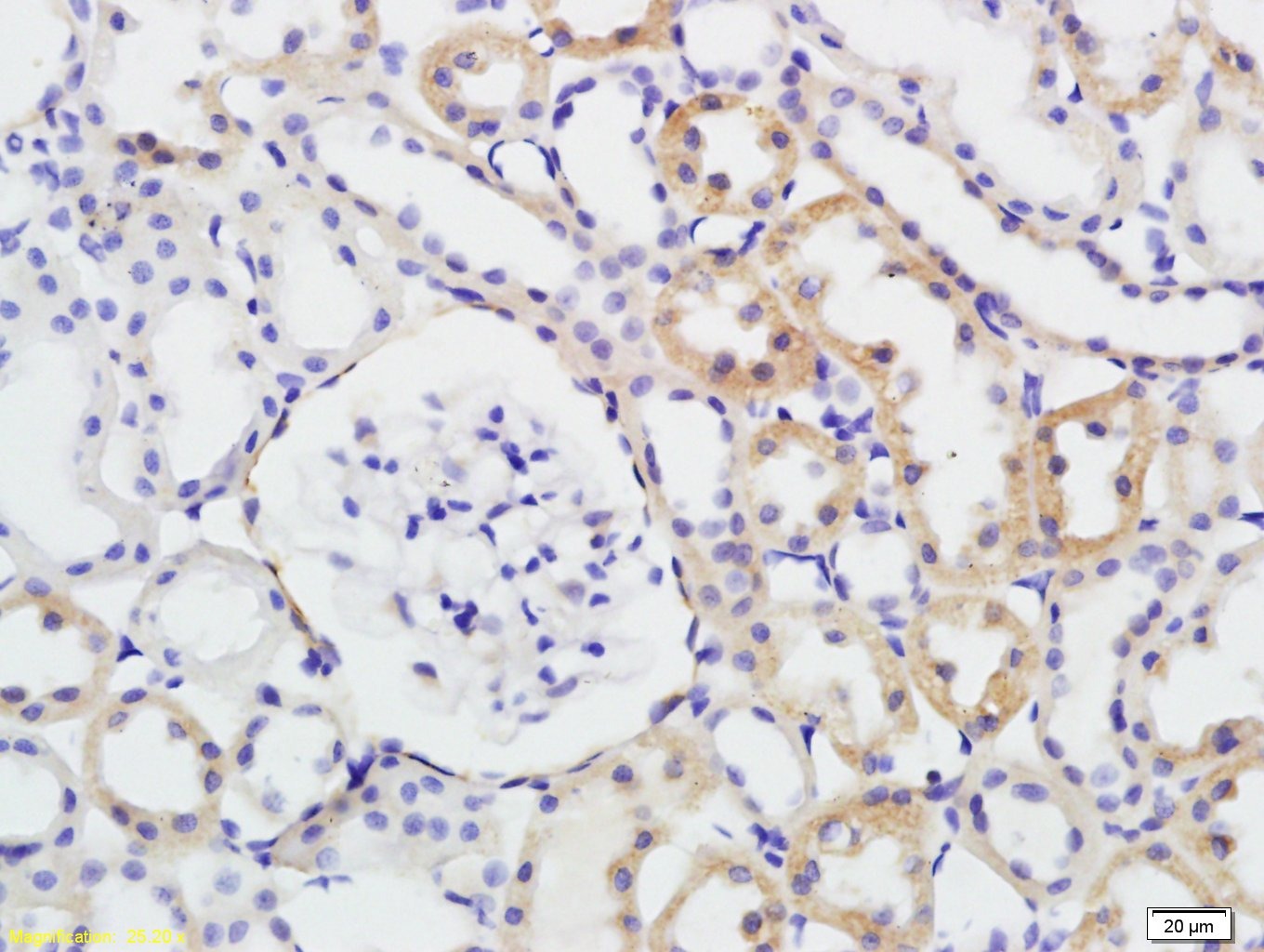 , ,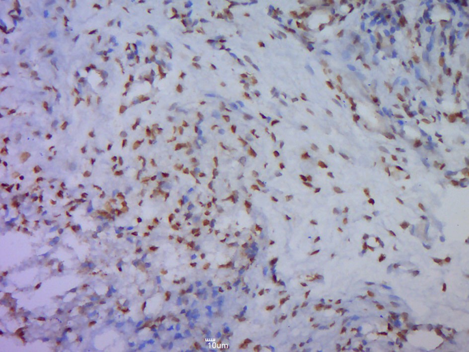 , ,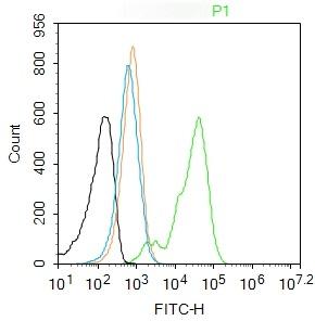 , ,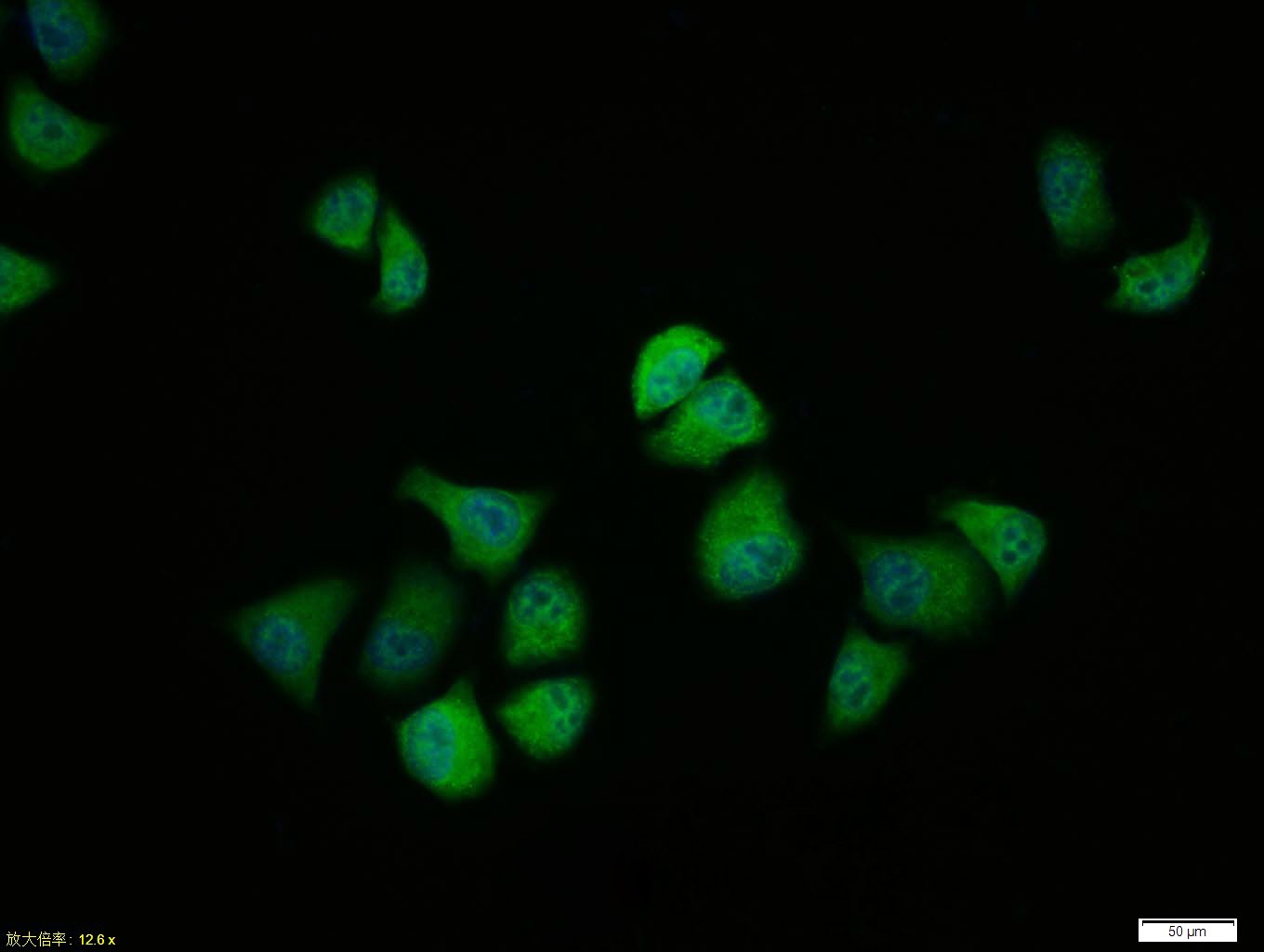 , ,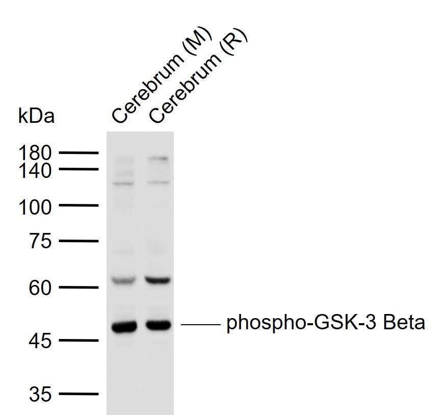 |
| Application | |
| Recommended Dose | FCM=1 μg/Test; ICC/IF=1:100-500; IF=1:100-500; IHC-Fr=1:100-500; IHC-P=1:100-500; WB=1:500-2000 |
| Antibody Type | Polyclonal |
| Host Species | Rabbit |
| Subcellular Localization | Cytoplasm. Nucleus. Cell membrane. Note=The phosphorylated form shows localization to cytoplasm and cell membrane. The MEMO1-RHOA-DIAPH1 signaling pathway controls localization of the phosophorylated form to the cell membrane. |
| Tissue Specificity | Expressed in testis, thymus, prostate and ovary and weakly expressed in lung, brain and kidney. |
| Construction | Polyclonal Antibody |
| Purification | Protein A purified |
| Appearance | Liquid |
| Formulation | 0.01M TBS (pH7.4) with 1% BSA, 0.02% Proclin300 and 50% Glycerol. |
| Concentration | 1 mg/mL |
| Research Background | The protein encoded by this gene is a serine-threonine kinase, belonging to the glycogen synthase kinase subfamily. It is involved in energy metabolism, neuronal cell development, and body pattern formation. Polymorphisms in this gene have been implicated in modifying risk of Parkinson disease, and studies in mice show that overexpression of this gene may be relevant to the pathogenesis of Alzheimer disease. Alternatively spliced transcript variants encoding different isoforms have been found for this gene.[provided by RefSeq, Sep 2009] |
| Immunogen | KLH conjugated Synthesised phosphopeptide: human GSK-3 Beta around the phosphorylation site of Ser9 |
| Antigen Species | Human |
| Gene Name | GSK3B |
| Gene ID | |
| Protein Name | Glycogen synthase kinase-3 beta |
| Uniprot ID | |
| Biology Area | Integration of energy metabolism,Metabolism of carbohydrates,Diabetes associated,Hypertrophy,Carbohydrate metabolism,Integration of energy,Cancer,Diabetes,Heart disease,Notch Pathway,Other Kinases,Cytoplasmic,Cytoplasmic |
| Function | Constitutively active protein kinase that acts as a negative regulator in the hormonal control of glucose homeostasis, Wnt signaling and regulation of transcription factors and microtubules, by phosphorylating and inactivating glycogen synthase (GYS1 or GYS2), EIF2B, CTNNB1/beta-catenin, APC, AXIN1, JUN, NFATC1/NFATC, MAPT/TAU and MACF1. Requires primed phosphorylation of the majority of its substrates. In skeletal muscle, contributes to insulin regulation of glycogen synthesis by phosphorylating and inhibiting GYS1 activity and hence glycogen synthesis. May also mediate the development of insulin resistance by regulating activation of transcription factors. Regulates protein synthesis by controlling the activity of initiation factor 2B (EIF2BE/EIF2B5) in the same manner as glycogen synthase. In Wnt signaling, GSK3B forms a multimeric complex with APC, AXIN1 and CTNNB1/beta-catenin and phosphorylates the N-terminus of CTNNB1 leading to its degradation mediated by ubiquitin/proteasomes. Phosphorylates JUN at sites proximal to its DNA-binding domain, thereby reducing its affinity for DNA. Phosphorylates NFATC1/NFATC on conserved serine residues promoting NFATC1/NFATC nuclear export, shutting off NFATC1/NFATC gene regulation, and thereby opposing the action of calcineurin. Phosphorylates MAPT/TAU on 'Thr-548', decreasing significantly MAPT/TAU ability to bind and stabilize microtubules. MAPT/TAU is the principal component of neurofibrillary tangles in Alzheimer disease. Plays an important role in ERBB2-dependent stabilization of microtubules at the cell cortex. Phosphorylates MACF1, inhibiting its binding to microtubules which is critical for its role in bulge stem cell migration and skin wound repair. Probably regulates NF-kappa-B (NFKB1) at the transcriptional level and is required for the NF-kappa-B-mediated anti-apoptotic response to TNF-alpha (TNF/TNFA). Negatively regulates replication in pancreatic beta-cells, resulting in apoptosis, loss of beta-cells and diabetes. Phosphorylates MUC1 in breast cancer cells, decreasing the interaction of MUC1 with CTNNB1/beta-catenin. Is necessary for the establishment of neuronal polarity and axon outgrowth. Phosphorylates MARK2, leading to inhibit its activity. Phosphorylates SIK1 at 'Thr-182', leading to sustain its activity. |
| Molecular Weight | Theoretical: 47 kDa. Actual: 47 kDa. |
| Stability & Storage | Store at -20°C or -80°C for 12 months. Avoid repeated freeze-thaw cycles. |
| Transport | Shipping with blue ice. |
| Size | Quantity | Unit Price | Amount | Operation |
|---|
 Hello! How can I help you today?
 Copyright © 2015-2026 TargetMol Chemicals Inc. All Rights Reserved.
