
Shopping Cart
Remove All Your shopping cart is currently empty

Synonyms: platelet-derived growth factor receptor, α polypeptide, platelet-derived growth factor receptor, alpha polypeptide
Anti-PDGFRA Polyclonal Antibody
| Pack Size | Price | USA Stock | Global Stock | Quantity |
|---|---|---|---|---|
| 50 µL | $221 | 7-10 days | 7-10 days | |
| 100 µL | $373 | 7-10 days | 7-10 days | |
| 200 µL | $528 | 7-10 days | 7-10 days |
| Description | Anti-PDGFRA Polyclonal Antibody is a Rabbit antibody targeting PDGFRA. Anti-PDGFRA Polyclonal Antibody can be used in FCM, IF, IHC-Fr, IHC-P. |
| Synonyms | platelet-derived growth factor receptor, α polypeptide, platelet-derived growth factor receptor, alpha polypeptide |
| Ig Type | IgG |
| Reactivity | Human,Mouse,Rat (predicted:Chicken,Dog,Pig,Cow,Horse) |
| Verified Activity | 1. Tissue/cell: human colon carcinoma; 4% Paraformaldehyde-fixed and paraffin-embedded; Antigen retrieval: citrate buffer (0.01M, pH6.0), Boiling bathing for 15 min; Block endogenous peroxidase by 3% Hydrogen peroxide for 30 min; Blocking buffer (normal goat serum) at 37°C for 20 min; Incubation: Anti-PDGFRA Polyclonal Antibody, Unconjugated (TMAB-01354) 1:200, overnight at 4°C, followed by conjugation to the secondary antibody and DAb staining. 2. Blank control: Mouse Kidney (blue). Primary Antibody: Rabbit Anti-PDGFRA antibody (TMAB-01354,Green); Dilution: 1 μg in 100 μL 1X PBS containing 0.5% BSA; Isotype Control Antibody: Rabbit Igg (orange),used under the same conditions; Secondary Antibody: Goat anti-rabbit IgG-FITC (white blue), Dilution: 1:200 in 1 X PBS containing 0.5% BSA. Protocol The cells were fixed with 2% paraformaldehyde for 10 min at 37°C. Primary antibody (TMAB-01354, 1 μg/1x10^6 cells) were incubated for 30 min at room temperature, followed by 1 X PBS containing 0.5% BSA + 10% goat serum (15 min) to block non-specific protein-protein interactions. Then the Goat Anti-rabbit IgG/FITC antibody was added into the blocking buffer mentioned above to react with the primary antibody at 1/200 dilution for 40 min on ice. 3. Black line: Positive blank control (SH-SY5Y); Negative blank control (A431) Green line: Primary Antibody (Rabbit Anti-PDGFRA antibody (TMAB-01354)) Orange line: Isotype Control Antibody (Rabbit IgG). Blue line: Secondary Antibody (Goat anti-rabbit IgG-AF488) SH-SY5Y (Positive) and A431 (Negative control) cells (black) were fixed with 4% PFA for 10 min at room temperature,permeabilized with 90% ice-cold methanol for 20 min at-20°C, and incubated in 5% BSA blocking buffer for 30 min at room temperature. Cells were then stained with PDGFRA Antibody (TMAB-01354) at 1:50 dilution in blocking buffer and incubated for 30 min at room temperature, washed twice with 2% BSA in PBS, followed by secondary antibody (blue) incubation for 40 min at room temperature. Acquisitions of 20,000 events were performed. Cells stained with primary antibody (green), and isotype control (orange). 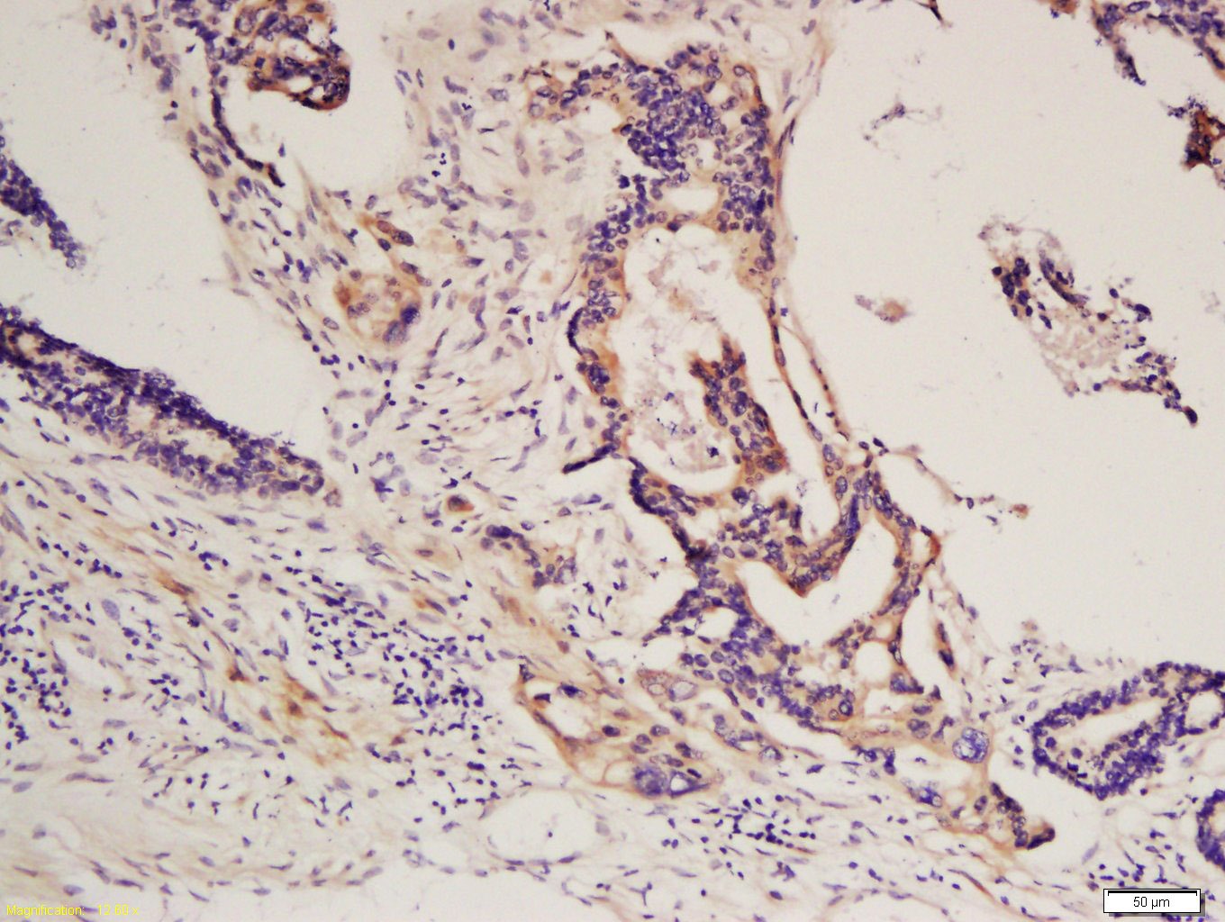 , , , ,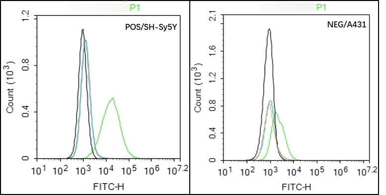 |
| Application | |
| Recommended Dose | IHC-P: 1:100-500; IHC-Fr: 1:100-500; IF: 1:100-500; FCM: 1μg/Test |
| Antibody Type | Polyclonal |
| Host Species | Rabbit |
| Subcellular Localization | Cell membrane; Single-pass type I membrane protein. Note=The activated receptor is rapidly internalized and degraded. |
| Tissue Specificity | Detected in platelets (at protein level). Widely expressed. Detected in brain, fibroblasts, smooth muscle, heart, and embryo. Expressed in primary and metastatic colon tumors and in normal colon tissue. |
| Construction | Polyclonal Antibody |
| Purification | Protein A purified |
| Appearance | Liquid |
| Formulation | 0.01M TBS (pH7.4) with 1% BSA, 0.02% Proclin300 and 50% Glycerol. |
| Concentration | 1 mg/mL |
| Research Background | This gene encodes a cell surface tyrosine kinase receptor for members of the platelet-derived growth factor family. These growth factors are mitogens for cells of mesenchymal origin. The identity of the growth factor bound to a receptor monomer determines whether the functional receptor is a homodimer or a heterodimer, composed of both platelet-derived growth factor receptor alpha and beta polypeptides. Studies suggest that this gene plays a role in organ development, wound healing, and tumor progression. Mutations in this gene have been associated with idiopathic hypereosinophilic syndrome, somatic and familial gastrointestinal stromal tumors, and a variety of other cancers. [provided by RefSeq, Mar 2012]. |
| Immunogen | KLH conjugated synthetic peptide: human PDGF-R-A |
| Antigen Species | Human |
| Gene Name | PDGFRA |
| Gene ID | |
| Protein Name | Platelet-derived growth factor receptor alpha |
| Uniprot ID | |
| Biology Area | PDGF,Growth factor receptors,Growth Factors,Inflammatory mediators,PDGF,Receptor Tyrosine Kinases |
| Function | Tyrosine-protein kinase that acts as a cell-surface receptor for PDGFA, PDGFB and PDGFC and plays an essential role in the regulation of embryonic development, cell proliferation, survival and chemotaxis. Depending on the context, promotes or inhibits cell proliferation and cell migration. Plays an important role in the differentiation of bone marrow-derived mesenchymal stem cells. Required for normal skeleton development and cephalic closure during embryonic development. Required for normal development of the mucosa lining the gastrointestinal tract, and for recruitment of mesenchymal cells and normal development of intestinal villi. Plays a role in cell migration and chemotaxis in wound healing. Plays a role in platelet activation, secretion of agonists from platelet granules, and in thrombin-induced platelet aggregation. Binding of its cognate ligands - homodimeric PDGFA, homodimeric PDGFB, heterodimers formed by PDGFA and PDGFB or homodimeric PDGFC -leads to the activation of several signaling cascades; the response depends on the nature of the bound ligand and is modulated by the formation of heterodimers between PDGFRA and PDGFRB. Phosphorylates PIK3R1, PLCG1, and PTPN11. Activation of PLCG1 leads to the production of the cellular signaling molecules diacylglycerol and inositol 1,4,5-trisphosphate, mobilization of cytosolic Ca(2+) and the activation of protein kinase C. Phosphorylates PIK3R1, the regulatory subunit of phosphatidylinositol 3-kinase, and thereby mediates activation of the AKT1 signaling pathway. Mediates activation of HRAS and of the MAP kinases MAPK1/ERK2 and/or MAPK3/ERK1. Promotes activation of STAT family members STAT1, STAT3 and STAT5A and/or STAT5B. Receptor signaling is down-regulated by protein phosphatases that dephosphorylate the receptor and its down-stream effectors, and by rapid internalization of the activated receptor. |
| Molecular Weight | Theoretical: 117 kDa. |
| Stability & Storage | Store at -20°C or -80°C for 12 months. Avoid repeated freeze-thaw cycles. |
| Transport | Shipping with blue ice. |
| Size | Quantity | Unit Price | Amount | Operation |
|---|
 Hello! How can I help you today?
 Copyright © 2015-2026 TargetMol Chemicals Inc. All Rights Reserved.
