
Shopping Cart
Remove All Your shopping cart is currently empty

Synonyms: Optic atrophy protein 1 homolog, Optic atrophy protein 1, Optic atrophy 1(autosomal dominant), Optic atrophy 1 homolog(human), Optic atrophy 1 gene protein, OPTIC ATROPHY 1, OPA1, OPA 1, OAK, NTG, NPG, Mitochondrial dynamin like GTPase, Mitochondrial dynamin like 120 kDa protein, MGM1, largeG, Large GTP binding protein, KJER type, KIAA0567, Juvenile kjer type optic atrophy, FLJ12460, Dynamin-like 120 kDa protein, form S1, Dynamin like 120 kDa protein, mitochondrial, Dynamin like 120 kDa protein
Anti-OPA1 Polyclonal Antibody
| Pack Size | Price | USA Stock | Global Stock | Quantity |
|---|---|---|---|---|
| 50 µL | $221 | 7-10 days | 7-10 days | |
| 100 µL | $373 | 7-10 days | 7-10 days | |
| 200 µL | $529 | 7-10 days | 7-10 days |
| Description | Anti-OPA1 Polyclonal Antibody is a Rabbit antibody targeting OPA1. Anti-OPA1 Polyclonal Antibody can be used in IF,IHC-Fr,IHC-P,WB. |
| Synonyms | Optic atrophy protein 1 homolog, Optic atrophy protein 1, Optic atrophy 1(autosomal dominant), Optic atrophy 1 homolog(human), Optic atrophy 1 gene protein, OPTIC ATROPHY 1, OPA1, OPA 1, OAK, NTG, NPG, Mitochondrial dynamin like GTPase, Mitochondrial dynamin like 120 kDa protein, MGM1, largeG, Large GTP binding protein, KJER type, KIAA0567, Juvenile kjer type optic atrophy, FLJ12460, Dynamin-like 120 kDa protein, form S1, Dynamin like 120 kDa protein, mitochondrial, Dynamin like 120 kDa protein |
| Ig Type | IgG |
| Reactivity | Rat (predicted:Human,Mouse,Dog,Pig,Cow,Horse,Rabbit,Sheep) |
| Verified Activity | 1. Tissue/cell: rat brain tissue; 4% Paraformaldehyde-fixed and paraffin-embedded; Antigen retrieval: citrate buffer (0.01M, pH6.0), Boiling bathing for 15 min; Block endogenous peroxidase by 3% Hydrogen peroxide for 30 min; Blocking buffer (normal goat serum) at 37°C for 20 min; Incubation: Anti-OPA1 Polyclonal Antibody, Unconjugated (TMAB-01288) 1:200, overnight at 4°C, followed by conjugation to the secondary antibody and DAb staining. 2. Sample: Cerebrum (Rat) Lysate at 40 μg Primary: Anti-OPA1 (TMAB-01288) at 1/2000 dilution Secondary: IRDye800CW Goat Anti-Rabbit IgG at 1/20000 dilution Predicted band size: 111 kDa Observed band size: 111 kDa 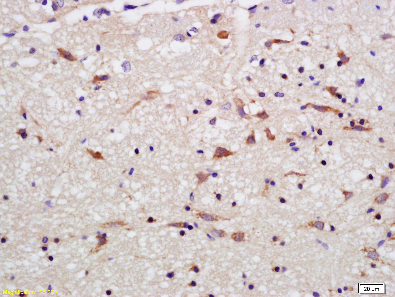 , ,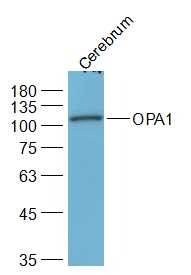 |
| Application | |
| Recommended Dose | WB: 1:500-2000; IHC-P: 1:100-500; IHC-Fr: 1:100-500; IF: 1:100-500 |
| Antibody Type | Polyclonal |
| Host Species | Rabbit |
| Subcellular Localization | Mitochondrion inner membrane. Mitochondrion intermembrane space. |
| Tissue Specificity | Highly expressed in retina. Also expressed in brain, testis, heart and skeletal muscle. Isoform 1 expressed in retina, skeletal muscle, heart, lung, ovary, colon, thyroid gland, leukocytes and fetal brain. Isoform 2 expressed in colon, liver, kidney, thyr |
| Construction | Polyclonal Antibody |
| Purification | Protein A purified |
| Appearance | Liquid |
| Formulation | 0.01M TBS (pH7.4) with 1% BSA, 0.02% Proclin300 and 50% Glycerol. |
| Concentration | 1 mg/mL |
| Research Background | OPA1 is a 120kDa protein belonging to the dynamin family. The OPA1 gene has been localized to 3q29. The gene is targeted to mitochondria and is involved in mitochondrial biogenesis. Defects in OPA1 are a cause of optic atrophy type 1. OPA1 is mostly expressed in retina but can also be expressed in brain, testis, heart and skeletal muscle. |
| Immunogen | KLH conjugated synthetic peptide: human OPA1 |
| Antigen Species | Human |
| Gene Name | OPA1 |
| Gene ID | |
| Protein Name | Dynamin-like 120 kDa protein, mitochondrial |
| Uniprot ID | |
| Biology Area | Cardiac metabolism,Mitophagy fission and fusion,Neurodegenerative disease,Visual system |
| Function | Dynamin-related GTPase required for mitochondrial fusion and regulation of apoptosis. May form a diffusion barrier for proteins stored in mitochondrial cristae. Proteolytic processing in response to intrinsic apoptotic signals may lead to disassembly of OPA1 oligomers and release of the caspase activator cytochrome C (CYCS) into the mitochondrial intermembrane space. |
| Molecular Weight | Theoretical: 111 kDa. Actual: 111 kDa. |
| Stability & Storage | Store at -20°C or -80°C for 12 months. Avoid repeated freeze-thaw cycles. |
| Transport | Shipping with blue ice. |
| Size | Quantity | Unit Price | Amount | Operation |
|---|
 Hello! How can I help you today?
 Copyright © 2015-2026 TargetMol Chemicals Inc. All Rights Reserved.
