Shopping Cart
Remove All Your shopping cart is currently empty
Anti-MAP1LC3A Antibody (4Q42) is a Rabbit antibody targeting MAP1LC3A. Anti-MAP1LC3A Antibody (4Q42) can be used in FCM,ICC/IF,IHC,IP,WB.
| Pack Size | Price | USA Warehouse | Global Warehouse | Quantity |
|---|---|---|---|---|
| 50 μL | $297 | 7-10 days | 7-10 days | |
| 100 μL | $498 | 7-10 days | 7-10 days |
| Description | Anti-MAP1LC3A Antibody (4Q42) is a Rabbit antibody targeting MAP1LC3A. Anti-MAP1LC3A Antibody (4Q42) can be used in FCM,ICC/IF,IHC,IP,WB. |
| Synonyms | microtubule-associated protein 1 light chain 3 α, microtubule-associated protein 1 light chain 3 alpha, MAP1BLC3, MAP1ALC3, LC3A, LC3, ATG8E |
| Ig Type | IgG |
| Clone | 4Q42 |
| Reactivity | Human,Mouse,Rat |
| Verified Activity | 1. Western blot analysis of MAP1LC3A on different lysates using anti-MAP1LC3A antibody at 1/1,000 dilution. Positive control: Lane 1: SHG-44, Lane 2: Mouse brain, Lane 3: Mouse liver, Lane 4: Mouse skeletal muscle. 2. Immunohistochemical analysis of paraffin-embedded human liver tissue using anti-MAP1LC3A antibody. Counter stained with hematoxylin. 3. Immunohistochemical analysis of paraffin-embedded mouse liver tissue using anti-MAP1LC3A antibody. Counter stained with hematoxylin. 4. Immunohistochemical analysis of paraffin-embedded mouse brain tissue using anti-MAP1LC3A antibody. Counter stained with hematoxylin. 5. ICC staining MAP1LC3A in Hela cells (green). The nuclear counter stain is DAPI (blue). Cells were fixed in paraformaldehyde, permeabilised with 0.25% Triton X100/PBS. 6. ICC staining MAP1LC3A in PC12 cells (green). The nuclear counter stain is DAPI (blue). Cells were fixed in paraformaldehyde, permeabilised with 0.25% Triton X100/PBS. 7. ICC staining MAP1LC3A in HUVEC cells (green). The nuclear counter stain is DAPI (blue). Cells were fixed in paraformaldehyde, permeabilised with 0.25% Triton X100/PBS. 8. Flow cytometric analysis of SH-SY-5Y cells with MAP1LC3A antibody at 1/50 dilution (blue) compared with an unlabelled control (cells without incubation with primary antibody; red). Alexa Fluor 488-conjugated goat anti rabbit IgG was used as the secondary antibody. 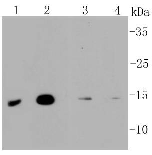 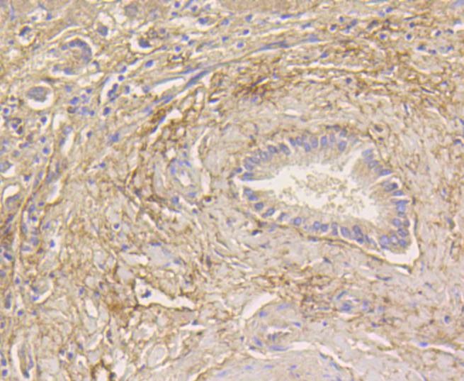 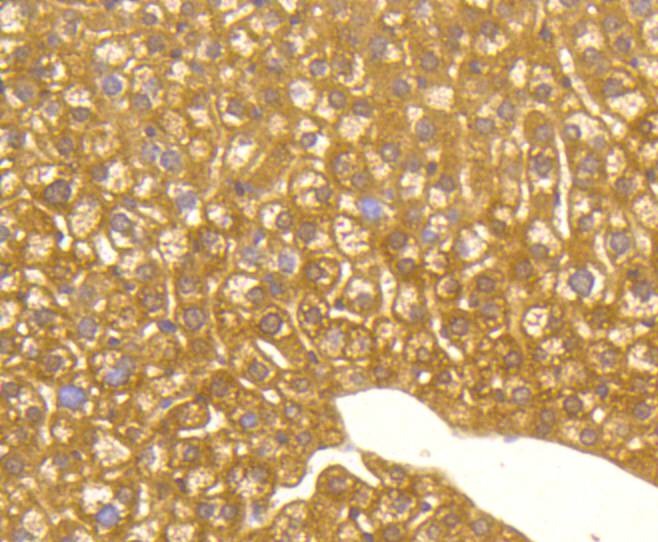 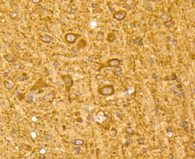 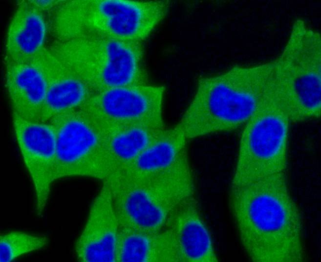 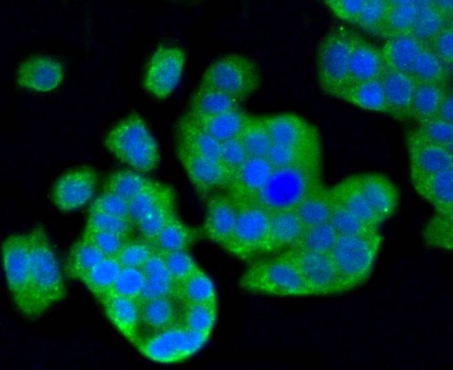 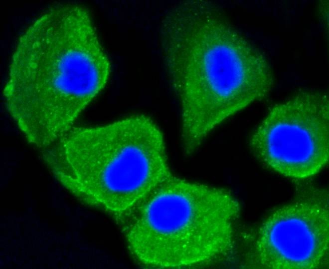 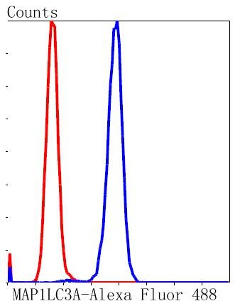 |
| Application | |
| Recommended Dose | WB: 1:1000-2000; IHC: 1:50-200; ICC/IF: 1:50-200; FCM: 1:50-100 |
| Antibody Type | Monoclonal |
| Host Species | Rabbit |
| Construction | Recombinant Antibody |
| Purification | ProA affinity purified |
| Appearance | Liquid |
| Formulation | 1*TBS (pH7.4), 1%BSA, 40%Glycerol. Preservative: 0.05% Sodium Azide. |
| Research Background | Microtubules, the primary component of the cytoskeletal network, interact with proteins called microtubule-associated proteins (MAPs). The microtubule-associated proteins can be divided into two groups, structural and dynamic. The structural microtubule-associated proteins, MAP-1A, MAP-1B, MAP-2A, MAP-2B and MAP-2C, stimulate tubulin assembly, enhance micro-tubule stability and influence the spatial distribution of microtubules within cells. Both MAP-1 and, to a greater extent, MAP-2 have been implicated as agents of microtubule depolymerization by suppressing the dynamic instability of the microtubules. The suppression of microtubule dynamic instability by the MAP proteins is thought to be associated with phosphorylation of the MAPs. |
| Conjucates | Unconjugated |
| Immunogen | Recombinant Protein |
| Uniprot ID |
| Molecular Weight | Theoretical: 14 kDa. |
| Stability & Storage | Store at -20°C or -80°C for 12 months. Avoid repeated freeze-thaw cycles. |
| Transport | Shipping with blue ice. |
| Size | Quantity | Unit Price | Amount | Operation |
|---|

Copyright © 2015-2026 TargetMol Chemicals Inc. All Rights Reserved.