
Shopping Cart
Remove All Your shopping cart is currently empty

Synonyms: XLKD1, LYVE-1, Lymphatic vessel endothelial hyaluronic acid receptor 1, Hyaluronic acid receptor, HAR, Extracellular link domain-containing protein 1, CRSBP-1, CRSBP1, Cell surface retention sequence-binding protein 1
Anti-LYVE1 Antibody
(7P115)
| Pack Size | Price | USA Stock | Global Stock | Quantity |
|---|---|---|---|---|
| 25 µL | $154 | 7-10 days | 7-10 days | |
| 50 µL | $273 | 7-10 days | 7-10 days | |
| 100 µL | $487 | 7-10 days | 7-10 days |
| Description | Anti-LYVE1 Antibody (7P115) is a Rabbit antibody targeting LYVE1. Anti-LYVE1 Antibody (7P115) can be used in WB. |
| Synonyms | XLKD1, LYVE-1, Lymphatic vessel endothelial hyaluronic acid receptor 1, Hyaluronic acid receptor, HAR, Extracellular link domain-containing protein 1, CRSBP-1, CRSBP1, Cell surface retention sequence-binding protein 1 |
| Ig Type | IgG |
| Clone | 7P115 |
| Reactivity | Mouse |
| Verified Activity | Sample: Lane 1: Mouse Spleen tissue lysates Lane 2: Mouse Blood cell lysates Lane 3: Mouse LympHnode tissue lysates Lane 4: Mouse Large intestine tissue lysates Lane 5: Rat Thyroid gland tissue lysates Lane 6: Rat Spleen tissue lysates Lane 7: Rat LympHnode tissue lysates Lane 8: Rat Large intestine tissue lysates Lane 9: Human Huvec cell lysates Lane 10: Human K562 cell lysates Primary: Anti-LYVE-1 (TMAB-01092) at 1/1000 dilution Secondary: IRDye800CW Goat Anti-Rabbit IgG at 1/20000 dilution Predicted band size: 32 kDa Observed band size: 58 kDa 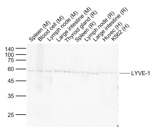 |
| Application | |
| Recommended Dose | WB=1:500-2000 |
| Antibody Type | Monoclonal |
| Host Species | Rabbit |
| Subcellular Localization | Membrane; Single-pass type I membrane protein. Note=Localized to the plasma membrane and in vesicles near extranuclear membranes which may represent trans-Golgi network (TGN) and endosomes/prelysosomeal compartments. Undergoes ligand-dependent internalization and recycling at the cell surface. |
| Tissue Specificity | Restricted to tissues or cell lines expressing erythroid characters. |
| Construction | Recombinant Antibody |
| Purification | Protein A purified |
| Appearance | Liquid |
| Formulation | 0.01M TBS (pH7.4) with 1% BSA, 0.02% Proclin300 and 50% Glycerol. |
| Concentration | 1 mg/mL |
| Research Background | The lymphatic vasculature forms a second circulatory system that drains extracellular fluid from the tissues and provides an exclusive environment in which immune cells can encounter and respond to foreign antigen. Recently a number of interesting molecules have been identified that may be exploited as markers for lymphatic endothelium, including the hyaluronan receptor LYVE1, PALE, VEGFR3, podoplanin. LYVE1 has been identified as a major receptor for HA (extracellular matrix glycosaminoglycan hyaluronan) on the lymph vessel wall. The deduced amino acid sequence of LYVE1 predicts a 322-residue type I integral membrane polypeptide 41% similar to the CD44 HA receptor with a 212-residue extracellular domain containing a single Link module the prototypic HA binding domain of the Link protein superfamily. Like CD44, the LYVE1 molecule binds both soluble and immobilized HA. However, unlike CD44, the LYVE1 molecule colocalizes with HA on the luminal face of the lymph vessel wall and is completely absent from blood vessels. Hence, LYVE1 is the first lymph-specific HA receptor to be characterized and is a uniquely powerful marker for lymph vessels themselves. |
| Immunogen | Recombinant Protein: mouse LYVE1 |
| Antigen Species | Mouse |
| Gene Name | LYVE1 |
| Gene ID | |
| Protein Name | Lymphatic vessel endothelial hyaluronic acid receptor 1 |
| Uniprot ID | |
| Biology Area | Endothelial Cell Markers,Endothelium |
| Function | Ligand-specific transporter trafficking between intracellular organelles (TGN) and the plasma membrane. Plays a role in autocrine regulation of cell growth mediated by growth regulators containing cell surface retention sequence binding (CRS). May act as an hyaluronan (HA) transporter, either mediating its uptake for catabolism within lymphatic endothelial cells themselves, or its transport into the lumen of afferent lymphatic vessels for subsequent re-uptake and degradation in lymph nodes. |
| Molecular Weight | Theoretical: 32 kDa. |
| Stability & Storage | Store at -20°C or -80°C for 12 months. Avoid repeated freeze-thaw cycles. |
| Transport | Shipping with blue ice. |
| Size | Quantity | Unit Price | Amount | Operation |
|---|
 Hello! How can I help you today?
 Copyright © 2015-2026 TargetMol Chemicals Inc. All Rights Reserved.
