Shopping Cart
Remove All Your shopping cart is currently empty
Anti-LGR-5 Polyclonal Antibody is a Rabbit antibody targeting LGR-5. Anti-LGR-5 Polyclonal Antibody can be used in FCM, IF, IHC-Fr, IHC-P, WB.
| Pack Size | Price | USA Warehouse | Global Warehouse | Quantity |
|---|---|---|---|---|
| 50 μL | $220 | 7-10 days | 7-10 days | |
| 100 μL | $372 | 7-10 days | 7-10 days | |
| 200 μL | $528 | 7-10 days | 7-10 days |
| Description | Anti-LGR-5 Polyclonal Antibody is a Rabbit antibody targeting LGR-5. Anti-LGR-5 Polyclonal Antibody can be used in FCM, IF, IHC-Fr, IHC-P, WB. |
| Synonyms | LGR5, HG38, G-protein coupled receptor HG38, G-protein coupled receptor 67, G-protein coupled receptor 49, GPR67, GPR49 |
| Ig Type | IgG |
| Reactivity | Human,Mouse (predicted:Rat) |
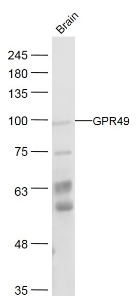
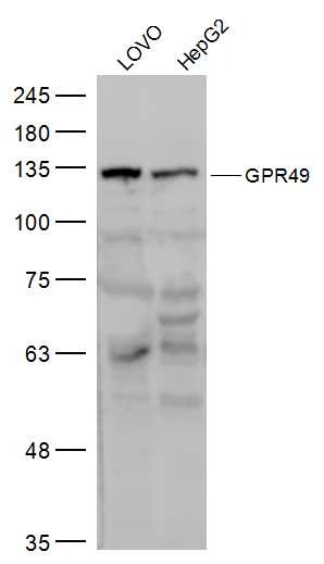
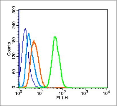
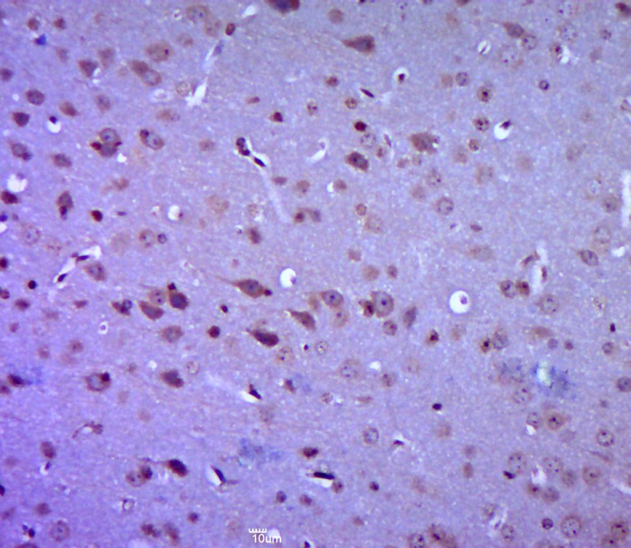
| Verified Activity | 1. Sample: Brain (Mouse) Lysate at 40 μg Primary: Anti-GPR49 (TMAB-01063) at 1/300 dilution Secondary: IRDye800CW Goat Anti-Rabbit IgG at 1/20000 dilution Predicted band size: 98 kDa Observed band size: 98 kDa 2. Sample: LOVO (Human) Cell Lysate at 30 μg HepG2 (Human) Cell Lysate at 30 μg Primary: Anti-GPR49 (TMAB-01063) at 1/500 dilution Secondary: IRDye800CW Goat Anti-Rabbit IgG at 1/20000 dilution Predicted band size: 98 kDa Observed band size: 125 kDa 3. Blank control (blue line): Hela (blue). Primary Antibody (green line): Rabbit Anti-GPR49 antibody (TMAB-01063), Dilution: 3 μg/10^6 cells; Isotype Control Antibody (orange line): Rabbit IgG. Secondary Antibody (white blue line): Goat anti-rabbit IgG-FITC Dilution: 1 μg/test. Protocol The cells were fixed with 70% ethanol (Overnight at 4°C) and then permeabilized with 90% ice-cold methanol for 30 min on ice. Cells stained with Primary Antibody for 30 min at room temperature. The cells were then incubated in 1 X PBS/2% BSA/10% goat serum to block non-specific protein-protein interactions followed by the antibody for 15 min at room temperature. The secondary antibody used for 40 min at room temperature. 4. Paraformaldehyde-fixed, paraffin embedded (mouse brain tissue); Antigen retrieval by boiling in sodium citrate buffer (pH6.0) for 15 min; Block endogenous peroxidase by 3% hydrogen peroxide for 20 min; Blocking buffer (normal goat serum) at 37°C for 30 min; Antibody incubation with (GPR49) Polyclonal Antibody, Unconjugated (TMAB-01063) at 1:400 overnight at 4°C, followed by a conjugated secondary for 20 min and DAB staining.     |
| Application | |
| Recommended Dose | WB: 1:500-2000; IHC-P: 1:100-500; IHC-Fr: 1:100-500; IF: 1:100-500; FCM: 1μg/Test |
| Antibody Type | Polyclonal |
| Host Species | Rabbit |
| Subcellular Localization | Cell membrane; Multi-pass membrane protein. |
| Tissue Specificity | Expressed in skeletal muscle, placenta, spinal cord, and various region of brain. Expressed at the base of crypts in colonic and small mucosa stem cells. In premalignant cancer expression is not restricted to the cript base. Overexpressed in cancers of th |
| Construction | Polyclonal Antibody |
| Purification | Protein A purified |
| Appearance | Liquid |
| Formulation | 0.01M TBS (pH7.4) with 1% BSA, 0.02% Proclin300 and 50% Glycerol. |
| Concentration | 1 mg/mL |
| Research Background | The protein encoded by this gene is a leucine-rich repeat-containing receptor (LGR) and member of the G protein-coupled, 7-transmembrane receptor (GPCR) superfamily. The encoded protein is a receptor for R-spondins and is involved in the canonical Wnt signaling pathway. This protein plays a role in the formation and maintenance of adult intestinal stem cells during postembryonic development. Several transcript variants encoding different isoforms have been found for this gene. [provided by RefSeq, Sep 2015] |
| Immunogen | KLH conjugated synthetic peptide: mouse GPR49 |
| Antigen Species | Mouse |
| Gene Name | LGR5 |
| Gene ID | |
| Protein Name | Leucine-rich repeat-containing G-protein coupled receptor 5 |
| Uniprot ID | |
| Biology Area | GPCR,GPCR,Surface Molecules |
| Function | Receptor for R-spondins that potentiates the canonical Wnt signaling pathway and acts as a stem cell marker of the intestinal epithelium and the hair follicle. Upon binding to R-spondins (RSPO1, RSPO2, RSPO3 or RSPO4), associates with phosphorylated LRP6 and frizzled receptors that are activated by extracellular Wnt receptors, triggering the canonical Wnt signaling pathway to increase expression of target genes. In contrast to classical G-protein coupled receptors, does not activate heterotrimeric G-proteins to transduce the signal. Involved in the development and/or maintenance of the adult intestinal stem cells during postembryonic development. |
| Molecular Weight | Theoretical: 98 kDa. |
| Stability & Storage | Store at -20°C or -80°C for 12 months. Avoid repeated freeze-thaw cycles. |
| Transport | Shipping with blue ice. |
| Size | Quantity | Unit Price | Amount | Operation |
|---|

Copyright © 2015-2026 TargetMol Chemicals Inc. All Rights Reserved.