
Shopping Cart
Remove All Your shopping cart is currently empty

Synonyms: Renal Carcinoma Antigen NY-REN-59, L-Lactate Dehydrogenase A Chain, LDH-M, LDH-A, LDHA, LDH Muscle Subunit, Cell Proliferation-Inducing Gene 19 Protein
Anti-LDHA Antibody
(4K270)
| Pack Size | Price | USA Stock | Global Stock | Quantity |
|---|---|---|---|---|
| 50 µL | $298 | 7-10 days | 7-10 days | |
| 100 µL | $498 | 7-10 days | 7-10 days |
| Description | Anti-LDHA Antibody (4K270) is a Rabbit antibody targeting LDHA. Anti-LDHA Antibody (4K270) can be used in FCM,ICC/IF,IHC,IP,WB. |
| Synonyms | Renal Carcinoma Antigen NY-REN-59, L-Lactate Dehydrogenase A Chain, LDH-M, LDH-A, LDHA, LDH Muscle Subunit, Cell Proliferation-Inducing Gene 19 Protein |
| Ig Type | IgG |
| Clone | 4K270 |
| Reactivity | Human,Mouse,Rat,zebrafish |
| Verified Activity | 1. Western blot analysis of Lactate Dehydrogenase on different lysates using anti-Lactate Dehydrogenase antibody at 1/1,000 dilution. Positive control: Lane 1: Hela, Lane 2: A549, Lane 3: MCF-7. 2. Western blot analysis of Lactate Dehydrogenase on hybrid fish (crucian-carp) brain tissue lysate using anti-Lactate Dehydrogenase antibody at 1/500 dilution. 3. Immunohistochemical analysis of paraffin-embedded human liver tissue using anti-Lactate Dehydrogenase antibody. Counter stained with hematoxylin. 4. Immunohistochemical analysis of paraffin-embedded human breast carcinoma tissue using anti-Lactate Dehydrogenase antibody. Counter stained with hematoxylin. 5. Immunohistochemical analysis of paraffin-embedded mouse liver tissue using anti-Lactate Dehydrogenase antibody. Counter stained with hematoxylin. 6. Immunohistochemical analysis of paraffin-embedded mouse testis tissue using anti-Lactate Dehydrogenase antibody. Counter stained with hematoxylin. 7. Immunohistochemical analysis of paraffin-embedded human liver cancer tissue using anti-Lactate Dehydrogenase antibody. Counter stained with hematoxylin. 8. Immunohistochemical analysis of paraffin-embedded mouse skeletal muscle tissue using anti-Lactate Dehydrogenase antibody. Counter stained with hematoxylin. 9. ICC staining Lactate Dehydrogenase in A549 cells (green). The nuclear counter stain is DAPI (blue). Cells were fixed in paraformaldehyde, permeabilised with 0.25% Triton X100/PBS. 10. ICC staining Lactate Dehydrogenase in A431 cells (green). The nuclear counter stain is DAPI (blue). Cells were fixed in paraformaldehyde, permeabilised with 0.25% Triton X100/PBS. 11. Flow cytometric analysis of Hela cells with Lactate Dehydrogenase antibody at 1/50 dilution (red) compared with an unlabelled control (cells without incubation with primary antibody; black). 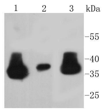 , ,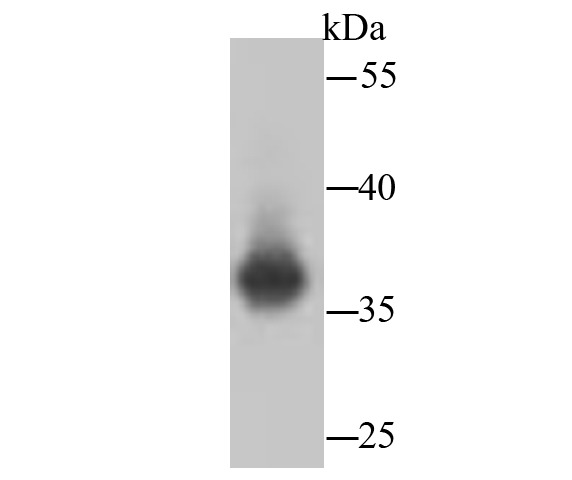 , ,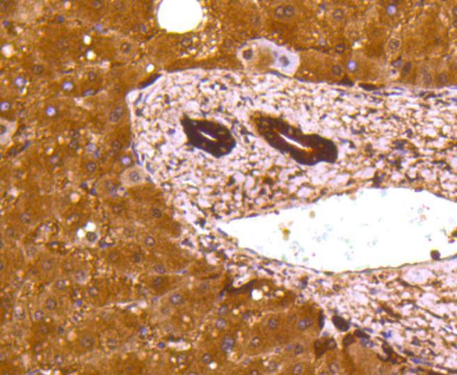 , ,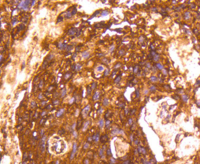 , ,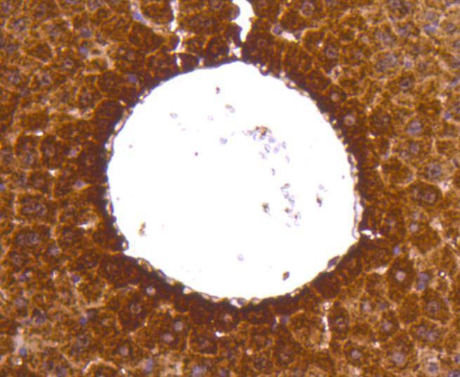 , ,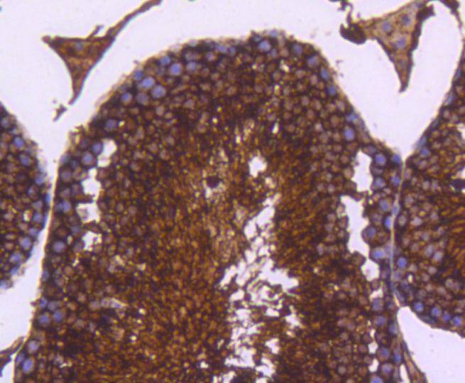 , ,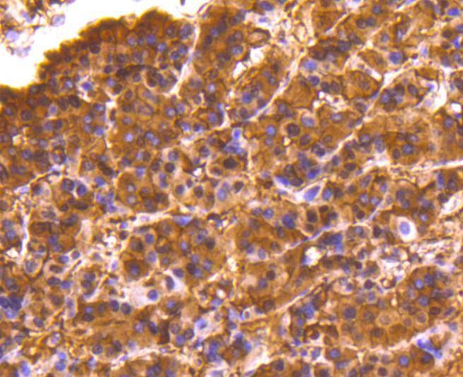 , ,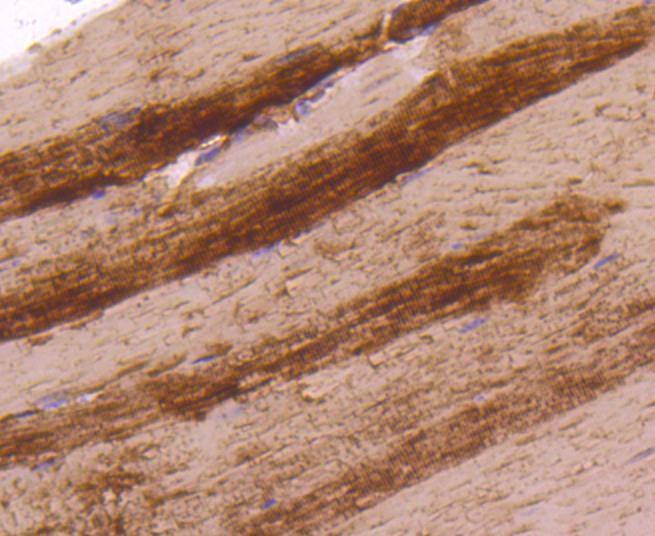 , ,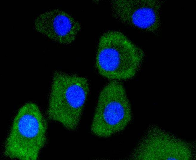 , ,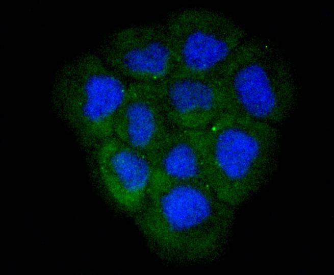 , ,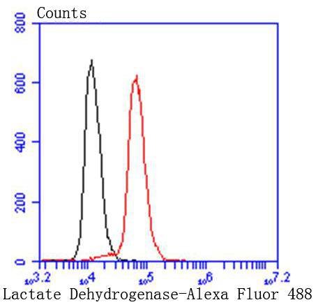 |
| Application | |
| Recommended Dose | WB: 1:1000-5000; IHC: 1:200-500; ICC/IF: 1:50-200; FCM: 1:50-100 |
| Antibody Type | Monoclonal |
| Host Species | Rabbit |
| Construction | Recombinant Antibody |
| Purification | ProA affinity purified |
| Appearance | Liquid |
| Formulation | 1*TBS (pH7.4), 1%BSA, 40%Glycerol. Preservative: 0.05% Sodium Azide. |
| Research Background | The lactate dehydrogenase family (LDH) catalyzes the final step of anaerobic glycolysis, the conversion of L-lactate and NAD to pyruvate and NADH. The LDH family consists of three members, LDH-A, LDH-B and LDH-C, all of which form tetramers consisting four subunits. However, each family member displays a specific tissue distribution pattern with LDH-A and LDH-B predominant in several tissues, specifically LDH-A in muscle and LDH-B in heart, while LDH-C expression is confined to the testis and sperm. LDHs function as powerful markers for germ cell tumors. The genes encoding human LDH-A and LDH-C map to chromosome 11, while the human LDH-B gene maps to chromosome 12. Deficiency in the LDH-A gene is linked to exertional myoglobinuria. |
| Conjucates | Unconjugated |
| Immunogen | Recombinant Protein |
| Uniprot ID |
| Molecular Weight | Theoretical: 37 kDa. |
| Stability & Storage | Store at -20°C or -80°C for 12 months. Avoid repeated freeze-thaw cycles. |
| Transport | Shipping with blue ice. |
| Size | Quantity | Unit Price | Amount | Operation |
|---|
 Hello! How can I help you today?
 Copyright © 2015-2026 TargetMol Chemicals Inc. All Rights Reserved.
