Shopping Cart
Remove All Your shopping cart is currently empty
Anti-Iba1 Antibody (1X229) is a Rabbit antibody targeting Iba1. Anti-Iba1 Antibody (1X229) can be used in FCM,ICC,IF,IHC,IP,WB.
| Pack Size | Price | USA Warehouse | Global Warehouse | Quantity |
|---|---|---|---|---|
| 50 μL | $296 | 7-10 days | 7-10 days | |
| 100 μL | $498 | 7-10 days | 7-10 days |
| Description | Anti-Iba1 Antibody (1X229) is a Rabbit antibody targeting Iba1. Anti-Iba1 Antibody (1X229) can be used in FCM,ICC,IF,IHC,IP,WB. |
| Synonyms | IRT-1, IRT1, IBA1, allograft inflammatory factor 1, AIF-1 |
| Ig Type | IgG |
| Clone | 1X229 |
| Reactivity | Human,Mouse,Rat |
| Verified Activity | 1. Immunohistochemical analysis of paraffin-embedded rat lung tissue using anti-Iba1 antibody. Counter stained with hematoxylin. 2. Immunohistochemical analysis of paraffin-embedded human lung cancer tissue using anti-Iba1 antibody. Counter stained with hematoxylin. 3. Immunohistochemical analysis of paraffin-embedded human spleen tissue using anti-Iba1 antibody. Counter stained with hematoxylin. 4. Immunohistochemical analysis of paraffin-embedded mouse brain tissue using anti-Iba1 antibody. Counter stained with hematoxylin. 5. Immunohistochemical analysis of paraffin-embedded mouse spleen tissue using anti-Iba1 antibody. Counter stained with hematoxylin. 6. ICC staining Iba1 in SH-SY5Y cells (green). The nuclear counter stain is DAPI (blue). Cells were fixed in paraformaldehyde, permeabilised with 0.25% Triton X100/PBS. 7. Flow cytometric analysis of THP-1 cells with Iba1 antibody at 1/100 dilution (red) compared with an unlabelled control (cells without incubation with primary antibody; black). 8. All lanes : Iba1 Antibody at 1/2k dilution, Lane 1: Mouse liver lysates, Lane 2: Mouse spleen lysates, Lane 3: Mouse lung lysates, Lane 4: Rat liver lysates, Lane 5: Rat spleen lysates, Lane 6: Rat lung lysates Lysates/proteins at 20 μg per lane. Secondary All lanes: Goat Anti-Rabbit IgG H&L (HRP) at 1/20000 dilution, Predicted band size: 17 kDa, Observed band size: 17 kDa, Exposure time: 120 seconds. 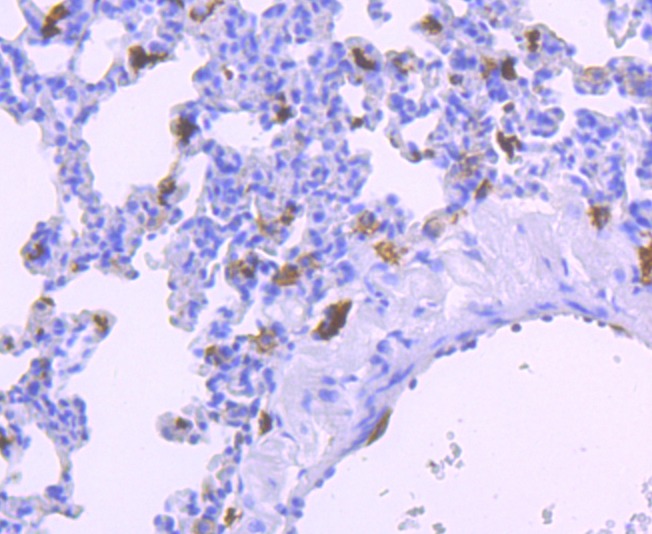 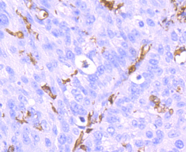 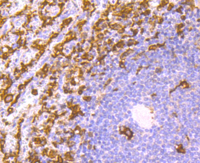 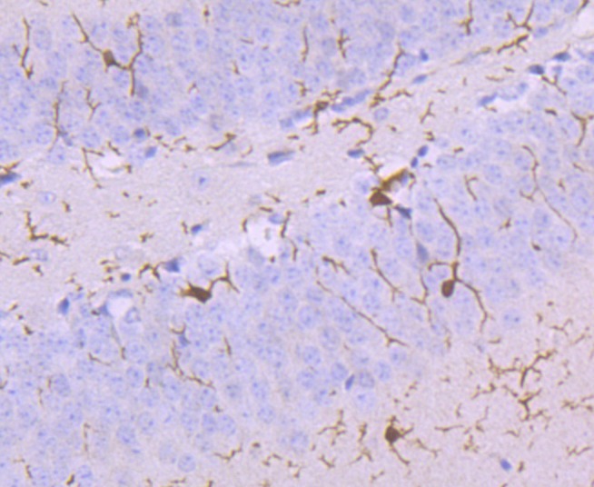 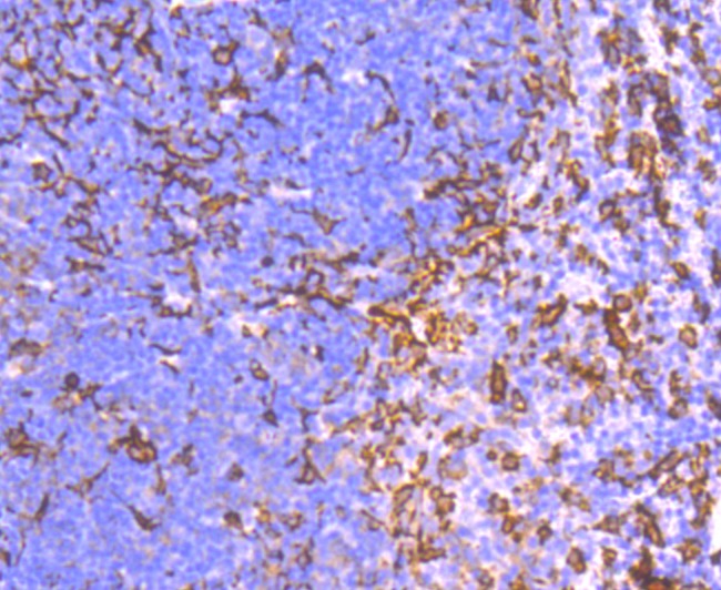 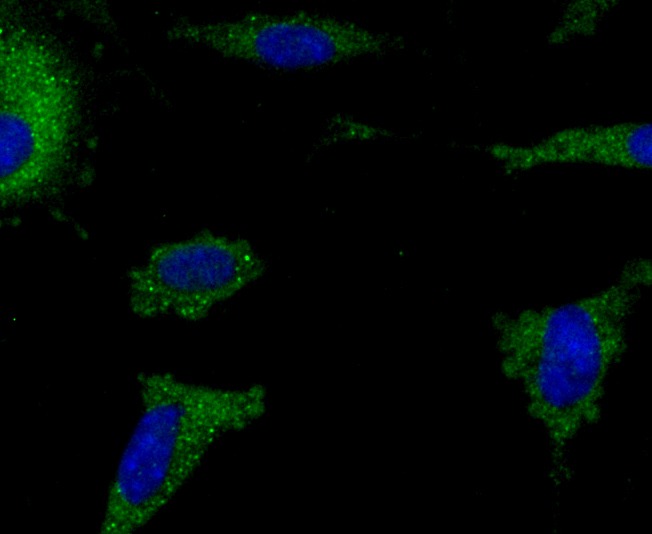 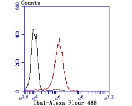 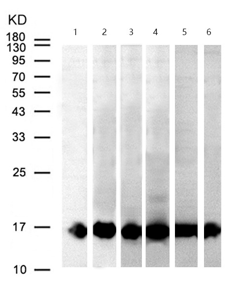 |
| Application | |
| Recommended Dose | WB: 1:500-1000; IHC: 1:100-500; ICC:IF: 1:50-100; IP: 1:10-50; FCM: 1:50-200 |
| Antibody Type | Monoclonal |
| Host Species | Rabbit |
| Construction | Recombinant Antibody |
| Purification | ProA affinity purified |
| Appearance | Liquid |
| Formulation | 1*TBS (pH7.4), 1%BSA, 40%Glycerol. Preservative: 0.05% Sodium Azide. |
| Research Background | Ionized calcium-binding adapter molecule 1 (Iba1), also known as allograft inflammatory factor-1 (AIF-1), is a 147 amino acid cytoplasmic, calcium-binding protein that is thought to play a role in macrophage activation and function. Iba1, containing two EF domains, is induced by cytokines and interferons. In an unstimulated state, Iba1 colocalizes with actin, and upon stimulation, translocates to lamellipodia. It is also a marker of human microglia and is expressed by macrophages in injured skeletal muscle. The gene encoding Iba1 maps to chromosome 6p21.33 and resides in the tumor necrosis factor (TNF) cluster of genes located in the region represented by the human major histocompatibility complex (MHC). |
| Conjucates | Unconjugated |
| Immunogen | Recombinant Protein |
| Uniprot ID |
| Molecular Weight | Theoretical: 17 kDa. |
| Stability & Storage | Store at -20°C or -80°C for 12 months. Avoid repeated freeze-thaw cycles. |
| Transport | Shipping with blue ice. |
| Size | Quantity | Unit Price | Amount | Operation |
|---|

Copyright © 2015-2026 TargetMol Chemicals Inc. All Rights Reserved.