
Shopping Cart
Remove All Your shopping cart is currently empty

Synonyms: RCCP2, MET proto-oncogene, receptor tyrosine kinase, HGFR, DFNB97, c-Met, AUTS9
Anti-HGFR/c-Met Polyclonal Antibody
| Pack Size | Price | USA Stock | Global Stock | Quantity |
|---|---|---|---|---|
| 50 µL | $220 | 7-10 days | 7-10 days | |
| 100 µL | $372 | 7-10 days | 7-10 days |
| Description | Anti-HGFR/c-Met Polyclonal Antibody is a Rabbit antibody targeting HGFR/c-Met. Anti-HGFR/c-Met Polyclonal Antibody can be used in ELISA,IF,IHC-Fr,IHC-P,WB. |
| Synonyms | RCCP2, MET proto-oncogene, receptor tyrosine kinase, HGFR, DFNB97, c-Met, AUTS9 |
| Ig Type | IgG |
| Reactivity | Human,Mouse,Rat |
| Verified Activity | 1. Sample: Lane 1: Mouse Kidney tissue lysates Lane 2: Mouse Stomach tissue lysates Lane 3: Mouse Lung tissue lysates Lane 4: Rat Kidney tissue lysates Lane 5: Rat Stomach tissue lysates Lane 6: Rat Lung tissue lysates Primary: Anti-MET (TMAB-00853) at 1/1000 dilution Secondary: IRDye800CW Goat Anti-Rabbit IgG at 1/20000 dilution Predicted band size: 33/123/153 kDa Observed band size: 145 kDa 2. Tissue/cell: human gastric carcinoma; 4% Paraformaldehyde-fixed and paraffin-embedded; Antigen retrieval: citrate buffer (0.01M, pH6.0), Boiling bathing for 15 min; Block endogenous peroxidase by 3% Hydrogen peroxide for 30 min; Blocking buffer (normal goat serum) at 37°C for 20 min; Incubation: Anti-C-Met Polyclonal Antibody, Unconjugated (TMAB-00853) 1:100, overnight at 4°C, followed by conjugation to the secondary antibody and DAb staining. 3. Tissue/cell: Human gastric cancer tissue; 4% Paraformaldehyde-fixed and paraffin-embedded; Antigen retrieval: citrate buffer (0.01M, pH6.0), Boiling bathing for 15 min; Block endogenous peroxidase by 3% Hydrogen peroxide for 30 min; Blocking buffer (normal goat serum) at 37°C for 20 min; Incubation: Anti-Met (c Met) Polyclonal Antibody, Unconjugated (TMAB-00853) 1:200, overnight at 4°C, followed by conjugation to the secondary antibody and DAb staining. 4. Tissue/cell: mouse intestine tissue;4% Paraformaldehyde-fixed and paraffin-embedded; Antigen retrieval: citrate buffer (0.01M, pH6.0), Boiling bathing for 15 min; Blocking buffer (normal goat serum) at 37°C for 20 min; Incubation: Anti-C-Met Polyclonal Antibody, Unconjugated (TMAB-00853) 1:200, overnight at 4°C; The secondary antibody was Goat Anti-Rabbit IgG, Cy3 conjugated used at 1:200 dilution for 40 minutes at 37°C. DAPI (5 μg/ml,blue) was used to stain the cell nucleus. 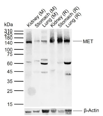 , ,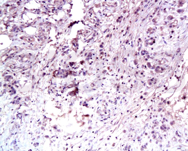 , ,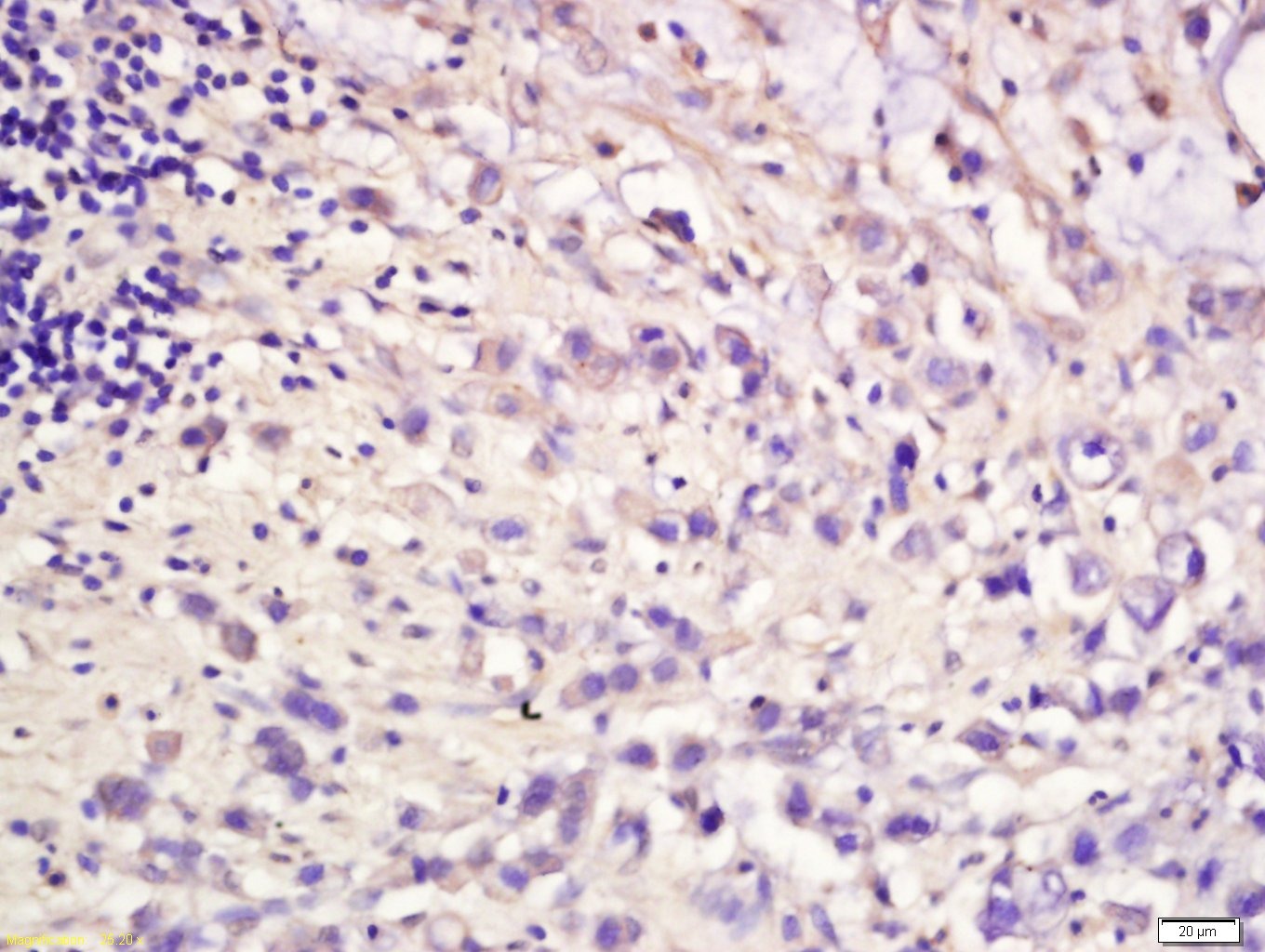 , ,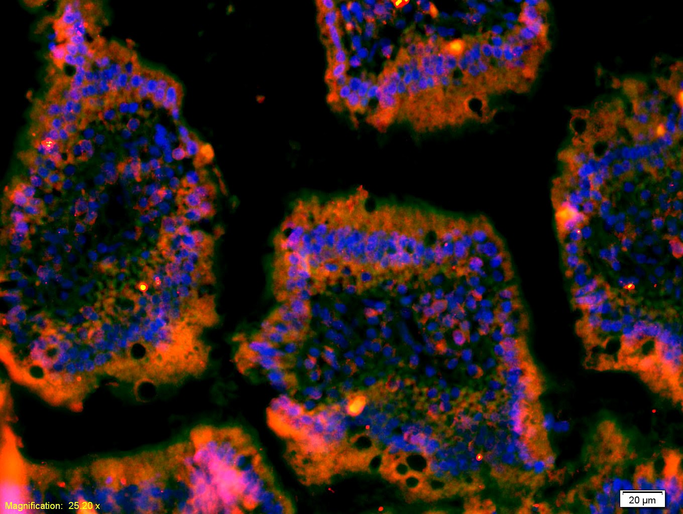 |
| Application | |
| Recommended Dose | WB: 1:500-2000; IHC-P: 1:100-500; IHC-Fr: 1:100-500; IF: 1:100-500; ELISA: 1:5000-10000 |
| Antibody Type | Polyclonal |
| Host Species | Rabbit |
| Subcellular Localization | Membrane; Single-pass type I membrane protein. Isoform 3: Secreted. |
| Tissue Specificity | Expressed in normal hepatocytes as well as in epithelial cells lining the stomach, the small and the large intestine. Found also in basal keratinocytes of esophagus and skin. High levels are found in liver, gastrointestinal tract, thyroid and kidney. Also |
| Construction | Polyclonal Antibody |
| Purification | Protein A purified |
| Appearance | Liquid |
| Formulation | 0.01M TBS (pH7.4) with 1% BSA, 0.02% Proclin300 and 50% Glycerol. |
| Concentration | 1 mg/mL |
| Research Background | This gene encodes a member of the receptor tyrosine kinase family of proteins and the product of the proto-oncogene MET. The encoded preproprotein is proteolytically processed to generate alpha and beta subunits that are linked via disulfide bonds to form the mature receptor. Further processing of the beta subunit results in the formation of the M10 peptide, which has been shown to reduce lung fibrosis. Binding of its ligand, hepatocyte growth factor, induces dimerization and activation of the receptor, which plays a role in cellular survival, embryogenesis, and cellular migration and invasion. Mutations in this gene are associated with papillary renal cell carcinoma, hepatocellular carcinoma, and various head and neck cancers. Amplification and overexpression of this gene are also associated with multiple human cancers. [provided by RefSeq, May 2016] |
| Immunogen | KLH conjugated synthetic peptide: mouse MET |
| Antigen Species | Mouse |
| Gene Name | MET |
| Gene ID | |
| Protein Name | Hepatocyte growth factor receptor |
| Uniprot ID | |
| Biology Area | Growth factor receptors,Proto-oncogenes,Receptor Tyrosine Kinases |
| Function | Receptor tyrosine kinase that transduces signals from the extracellular matrix into the cytoplasm by binding to hepatocyte growth factor/HGF ligand. Regulates many physiological processes including proliferation, scattering, morphogenesis and survival. Ligand binding at the cell surface induces autophosphorylation of MET on its intracellular domain that provides docking sites for downstream signaling molecules. Following activation by ligand, interacts with the PI3-kinase subunit PIK3R1, PLCG1, SRC, GRB2, STAT3 or the adapter GAB1. Recruitment of these downstream effectors by MET leads to the activation of several signaling cascades including the RAS-ERK, PI3 kinase-AKT, or PLCgamma-PKC. The RAS-ERK activation is associated with the morphogenetic effects while PI3K/AKT coordinates prosurvival effects. During embryonic development, MET signaling plays a role in gastrulation, development and migration of muscles and neuronal precursors, angiogenesis and kidney formation. In adults, participates in wound healing as well as organ regeneration and tissue remodeling. Promotes also differentiation and proliferation of hematopoietic cells.Acts as a receptor for Listeria internalin inlB, mediating entry of the pathogen into cells. |
| Molecular Weight | Theoretical: 33/123/156 kDa. Actual: 145 kDa. |
| Stability & Storage | Store at -20°C or -80°C for 12 months. Avoid repeated freeze-thaw cycles. |
| Transport | Shipping with blue ice. |
| Size | Quantity | Unit Price | Amount | Operation |
|---|
 Hello! How can I help you today?
 Copyright © 2015-2026 TargetMol Chemicals Inc. All Rights Reserved.
